. 2020 Jul;13(7):100769.
doi: 10.1016/j.tranon.2020.100769. Epub 2020 May 15.

MK2206 Enhances Cisplatin-Induced Cytotoxicity and Apoptosis in Testicular Cancer Through Akt Signaling Pathway Inhibition

Affiliations

MK2206 Enhances Cisplatin-Induced Cytotoxicity and Apoptosis in Testicular Cancer Through Akt Signaling Pathway Inhibition

Dingqi Sun et al. Transl Oncol. 2020 Jul.

Erratum in

Abstract

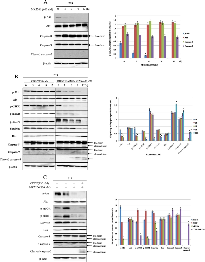
Objective: To improve conventional chemotherapeutic efficacy, it is significant to identify novel molecular markers for chemosensitivity as well as possible molecules accelerating cell-killing mechanisms. In this study, we attempted to elucidate how MK2206, an allosteric Akt inhibitor, enhances the cisplatin (CDDP)-induced cytotoxicity and apoptosis in testicular cancer.

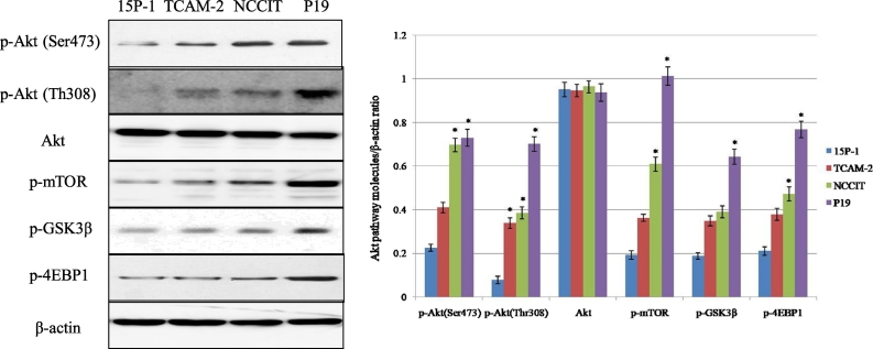
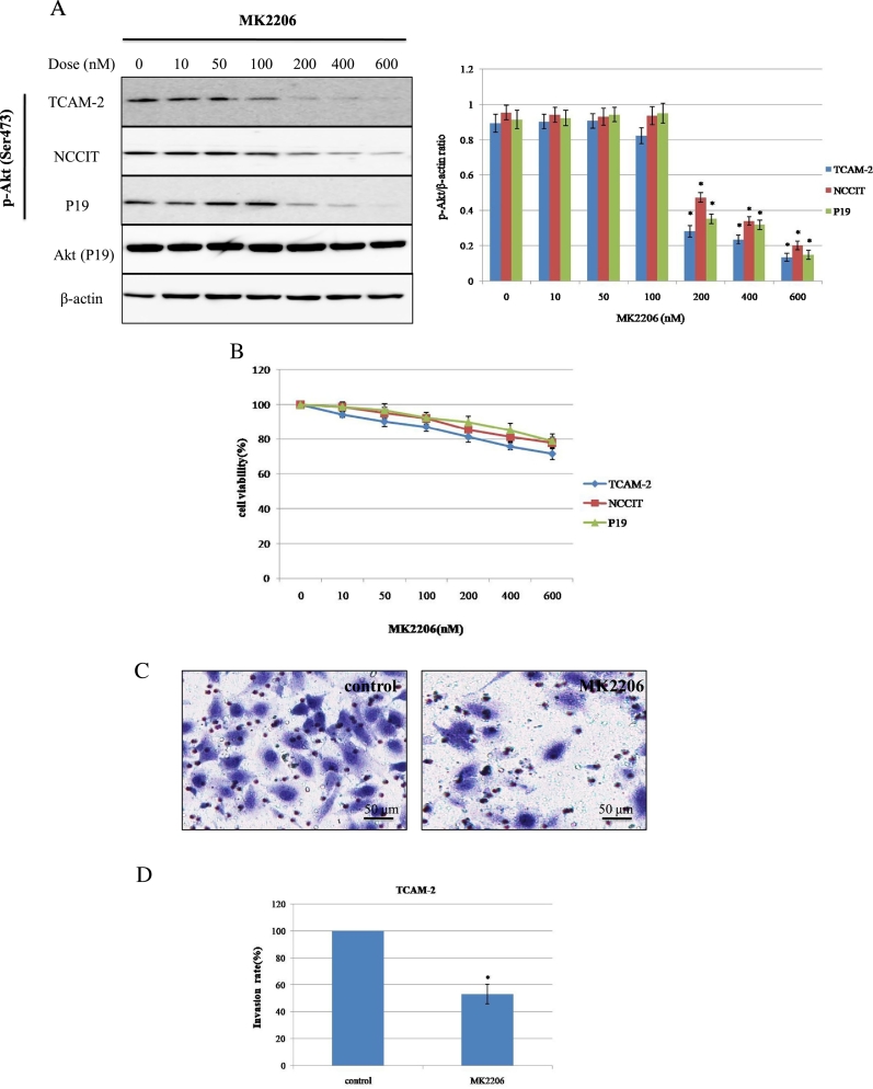
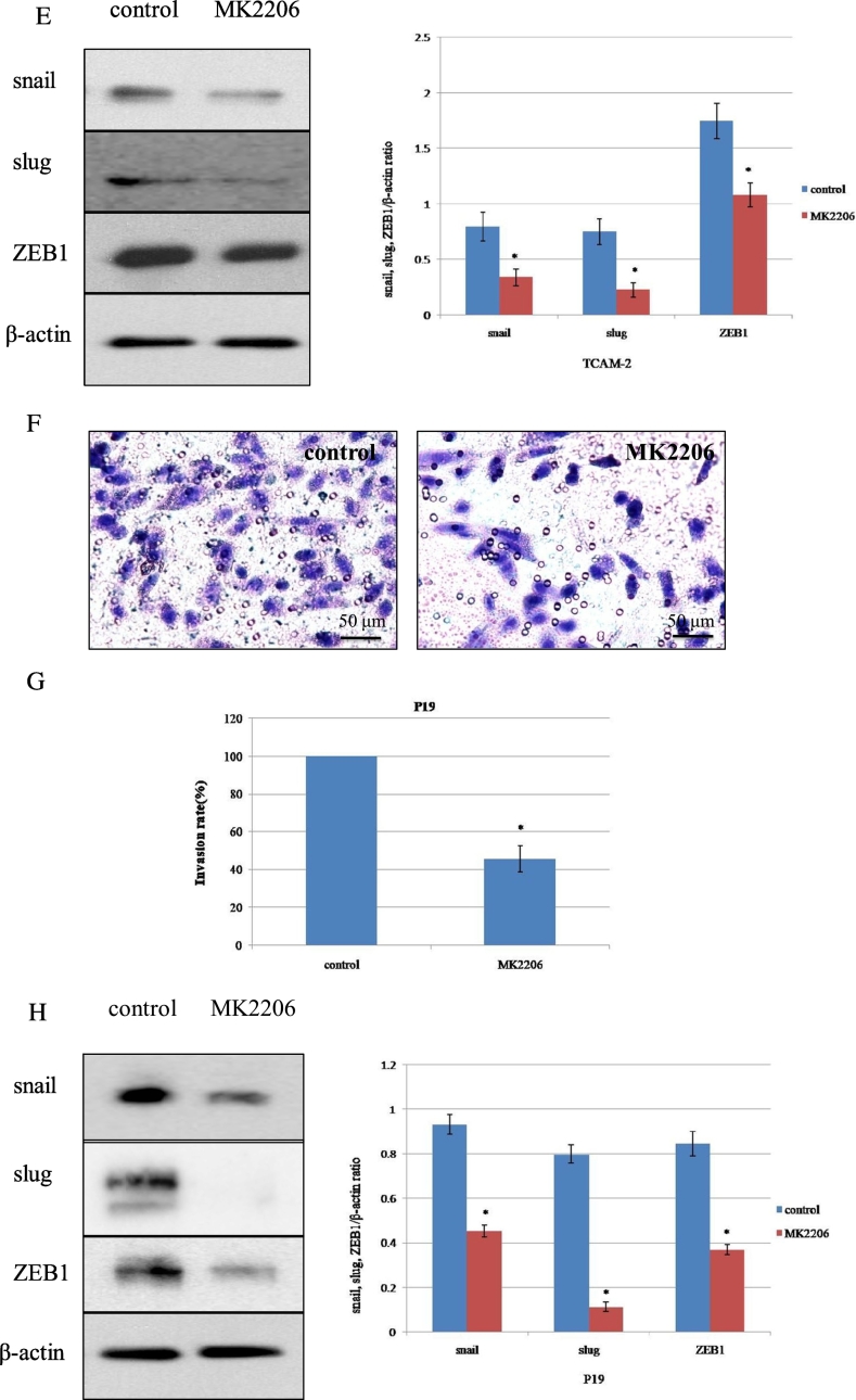
Materials and methods: We checked three testicular cancer cell lines for the expression of phospho(p)-Akt and its downstream molecules targets by Western blot. The potential antitumor effects were analyzed by MTT assay in vitro and by subcutaneous xenograft models in vivo. The cell invasion was analyzed by transwell invasion assay, and the activities of Akt signaling pathway and expression of apoptosis-related proteins were measured by Western blot.

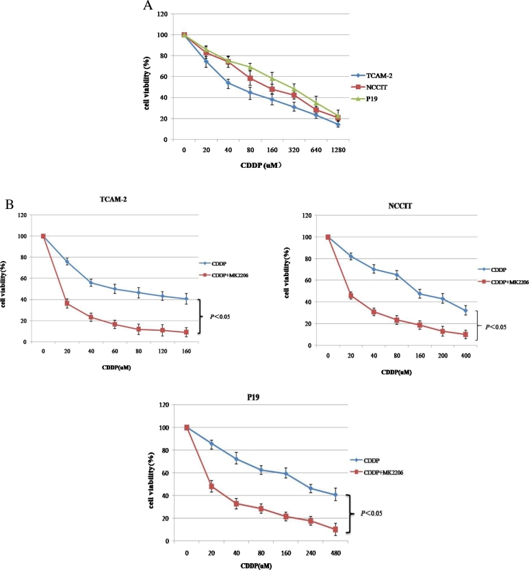
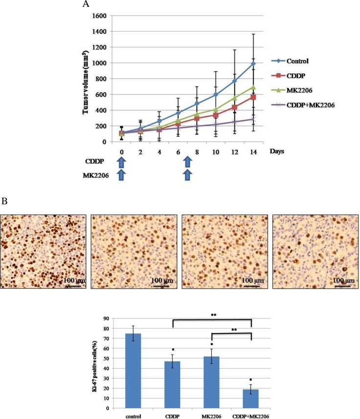
Results: Our results indicated that there was overactivation of p-Akt and its downstream molecules in testicular cancer cell lines compared with normal testis epithelium cells. MK2206 (600 nM) inhibited cell invasion in TCAM-2 and P19 cell lines and significantly increased the susceptibility of testicular cancer to CDDP. Combined with CDDP, MK2206 potentiated CDDP-induced cytotoxicity and apoptosis, with repressed expression of p-Akt and its downstream targets. The subcutaneous xenograft models also showed that a combined CDDP/MK2206 therapy completely suppressed tumor growth without any side effects.

Conclusion: These results suggested that the concomitant use of MK2206 could enhance the CDDP-induced cytotoxicity and apoptosis in testicular cancer with the suppressed expression of Akt pathway.


Figures

Figure 1
Figure 1
The expression status of the Akt signaling pathway in testicular cancer cell lines and one testicular epithelial cell line. Western blot shows the expression of Akt and its downstream molecular targets. Bars represent SD. *P < .05 vs. 15P-1.
Figure 2
Figure 2
The effect of MK2206 treatment on p-Akt expression, cell viability, and cell invasion in testicular cancer cell lines. (A) Cell lines were exposed to MK2206 at the indicated dose for 24 hours, and the expression level of p-Akt was examined using Western blot. The Akt expression of P19 cell line was shown. *P < .05 vs. 100 nM. (B) Cell viability curves after treatment with MK2206 as in (A). Mean values from at least 3 independent experiments are shown; bars represent SD. (C-D and F-G) The invasive capability of TCAM-2 and P19 were evaluated by transwell invasion assay. The cell suspension (5×10⁴ cells) and reagents (DMSO or MK2206) were added to the insert of a transwell cell culture chamber and incubated for 24 hours with medium containing 10% FBS in the bottom of the chamber. The cells that attached to the lower side of the membrane were fixed with 70% ethanol and stained with hematoxylin. Each experiment was triplicated, and the average number of cells in 5 microscopic fields (×200) was defined as the number of invasive cells. Representative microscopic image (C and F) and quantitated cell numbers (mean ± SD) of 3 independent experiments (D and G) were shown. (E and H) TCAM-2 and P19 cells were treated with control (DMSO) or MK2206 (600 nM) for 24 hours, and the abundance of indicated proteins related with invasion was examined using Western blot. SD = standard deviation. *P < .05 vs. control.
Figure 2
Figure 2
The effect of MK2206 treatment on p-Akt expression, cell viability, and cell invasion in testicular cancer cell lines. (A) Cell lines were exposed to MK2206 at the indicated dose for 24 hours, and the expression level of p-Akt was examined using Western blot. The Akt expression of P19 cell line was shown. *P < .05 vs. 100 nM. (B) Cell viability curves after treatment with MK2206 as in (A). Mean values from at least 3 independent experiments are shown; bars represent SD. (C-D and F-G) The invasive capability of TCAM-2 and P19 were evaluated by transwell invasion assay. The cell suspension (5×10⁴ cells) and reagents (DMSO or MK2206) were added to the insert of a transwell cell culture chamber and incubated for 24 hours with medium containing 10% FBS in the bottom of the chamber. The cells that attached to the lower side of the membrane were fixed with 70% ethanol and stained with hematoxylin. Each experiment was triplicated, and the average number of cells in 5 microscopic fields (×200) was defined as the number of invasive cells. Representative microscopic image (C and F) and quantitated cell numbers (mean ± SD) of 3 independent experiments (D and G) were shown. (E and H) TCAM-2 and P19 cells were treated with control (DMSO) or MK2206 (600 nM) for 24 hours, and the abundance of indicated proteins related with invasion was examined using Western blot. SD = standard deviation. *P < .05 vs. control.
Figure 3
Figure 3
The effect of combinatorial treatment with MK2206 and CDDP in testicular cancer cell lines. (A) Cell viability was evaluated 24 hours after treatment with CDDP at indicated concentrations using MTT assay among testicular cancer cell lines. (B) MTT assay showing effect of CDDP treatment with or without MK2206 (600 nM) in indicated cell lines. Average values from at least 3 independent experiments are shown; bars represent SD.
Figure 4
Figure 4
MK2206 enhances CDDP-induced apoptosis through the suppression of Akt pathway activity in testicular cancer cell lines. (A) Cells were exposed to MK2206 (600 nM) alone for the indicated times. “0 hours” refers to the time for addition of reagents. *P < .05 vs. 0 hours. (B) Cells were treated with CDDP at a concentration corresponding to IC30 (130 μM), with or without addition of 600 nM of MK2206 for the indicated times, and the abundance of indicated protein was examined using Western blot. *P < 0.05 vs. 0 hours. (C) Cells were harvested after treatment with or without CDDP at indicated doses corresponding to IC30 for each cell line in the presence or absence of 600 nM of MK2206 for 12 hours. Bars represent SD. *P < 0.05 vs. DMSO or CDDP.
Figure 5
Figure 5
The effect of combinatorial treatment with CDDP and MK2206 on the growth of a P19 subcutaneous xenograft tumor in vivo. (A) The change of subcutaneous P19 tumor volume measured using the Vernier caliper. After tumors reached the indicated volume, tumor-bearing mice were treated with indicated drugs as shown in the figure and described in section “Materials and methods”, and the effect of the indicated drugs on tumor growth was measured using the Vernier caliper. (B) Histological examination of the effect of CDDP and MK2206 on a P19 tumor in vivo. At day 15, the mice were sacrificed and the tumors were dissected. The expression of Ki-67 was assessed by immunohistochemistry. The percentage of Ki-67-positive cells in each treatment group was compared (*P < 0.05 vs. control; **P < 0.05, CDDP+MK2206 vs. CDDP; CDDP+ MK2206 vs. MK2206).

References

    1. Chia V.M., Li Y., Quraishi S.M. Effect modification of endocrine disruptors and testicular germ cell tumor risk by hormone-metabolizing genes. Int J Androl. 2010;33:588–596. - PMC - PubMed
    1. Datta S.R., Brunet A., Greenberg M.E. Cellular survival: a play in three Akts. Genes Dev. 1999;13:2905–2927. - PubMed
    1. Sun Y., Zhao S., Tian H. Depletion of PI3K p85alpha induces cell cycle arrest and apoptosis in colorectal cancer cells. Oncol Rep. 2009;22:1435–1441. - PubMed
    1. Liu N., Rowley B.R., Bull C.O. BAY 80-6946 is a highly selective intravenous PI3K inhibitor with potent p110α and p110δ activities in tumor cell lines and xenograft models. Mol Cancer Ther. 2013;12:2319–2330. - PubMed
    1. Guerrero-Zotano A., Mayer I.A., Arteaga C.L. PI3K/AKT/mTOR: role in breast cancer progression, drug resistance, and treatment. Cancer Metastasis Rev. 2016;35:515–552. - PubMed