. 2020 May 14;8(2):220.
doi: 10.3390/vaccines8020220.

A Novel Synonymous Mutation of SARS-CoV-2: Is This Possible to Affect Their Antigenicity and Immunogenicity?

Affiliations

A Novel Synonymous Mutation of SARS-CoV-2: Is This Possible to Affect Their Antigenicity and Immunogenicity?

Sung-Jae Kim et al. Vaccines (Basel). .

Abstract

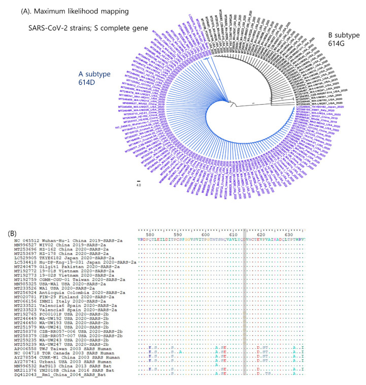
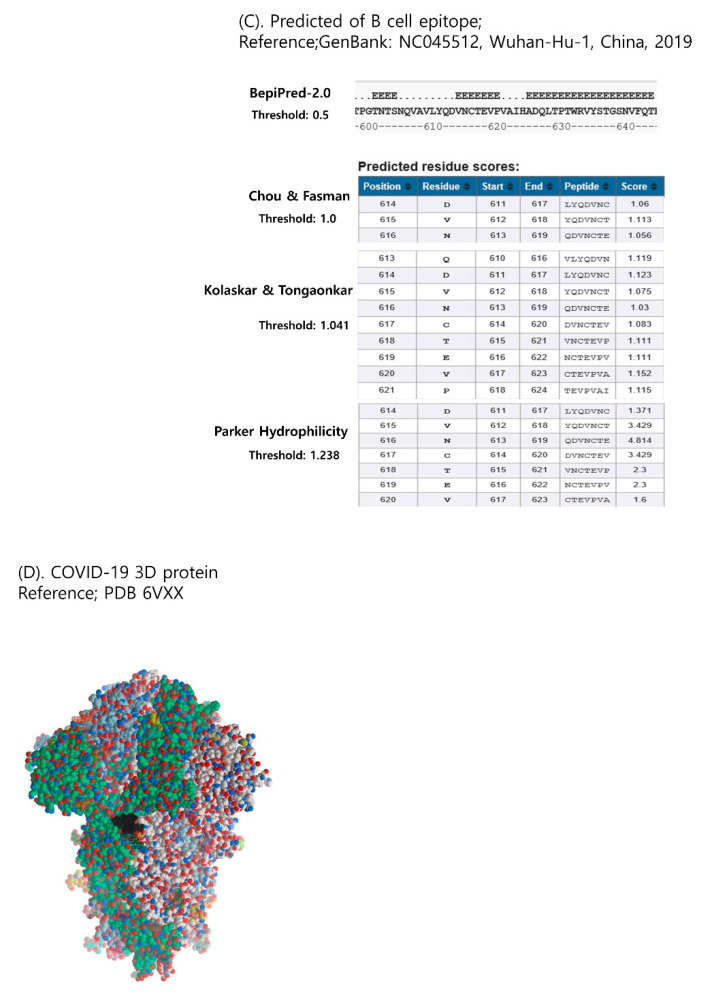
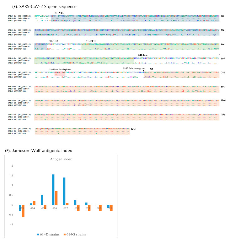
The S glycoprotein of coronaviruses is important for viral entry and pathogenesis with most variable sequences. Therefore, we analyzed the S gene sequences of SARS-CoV-2 to better understand the antigenicity and immunogenicity of this virus in this study. In phylogenetic analysis, two subtypes (SARS-CoV-2a and -b) were confirmed within SARS-CoV-2 strains. These two subtypes were divided by a novel synonymous mutation of D614G. This may play a crucial role in the evolution of SARS-CoV-2 to evade the host immune system. The region containing this mutation point was confirmed as a B-cell epitope located in the S1 domain, and SARS-CoV-2b strains exhibited severe reduced antigenic indexes compared to SARS-CoV-2a in this area. This may allow these two subtypes to have different antigenicity. If the two subtypes have different serological characteristics, a vaccine for both subtypes will be more effective to prevent COVID-19. Thus, further study is urgently required to confirm the antigenicity of these two subtypes.

Keywords: COVID-19; SARS-CoV-2; antigenicity; spike protein.


Conflict of interest statement

The authors declare that they have no competing interests.

Figures

Figure 1
Figure 1
Characterization of the complete S gene in SARS-CoV-2. (A) Phylogenetic analysis of SARS-CoV-2 strains based on the S gene. The phylogenetic trees were reconstructed from 144 sequences of SARS-CoV-2 collected globally. Thus, the two subtypes (SARS-CoV-2a and -2b) were completely divided. (B) Alignment of SARS-CoV sequences including the aa 614 position are highlighted in gray. A novel reliable synonymous mutation was identified to distinguish the A and B subtypes. SARS-Cov-2a and -2b strains consistently exhibited Ala (D) and Gly (G) at the amino acid sequence position 614, respectively. (C) Identification of B-cell epitopes in the adjacent area with aa 614. The B-cell epitope was predicted by BepiPred-2.0 [24], the Chou & Fasman method [25], the Kolaskar and Tongaonkar method [26], and Parker’s Hydrophilicity [27]. The 614–621 region was predicted to consist of epitopes. (D) The 3D-structure of SARS-CoV-2 spike protein by Mol soft Mol Browser 3.8–5 according to the original publication from the National Center for Biotechnology Information (NCBI): PDB;6VXX. The predicted B-cell epitope (aa 613–620) highlighted in black color was located at a relatively well-exposed part. (E) Sequence alignment of SARS-CoV-1 and -2. The S1 subunit is responsible for receptor binding and the S2 subunit mediates membrane fusion. The S1 subunit consists of two major domains capable of binding to host receptors: an amino (N)-terminal domain (NTD) and a carboxy (C)-terminal domain (CTD) and two sub-domains that may allow receptor-induced conformational changes: SD-1 and SD-2. (F) The antigenic index of each amino acid constituting this region (amino acids 613–621) by the Jameson–Wolf method [28].
Figure 1
Figure 1
Characterization of the complete S gene in SARS-CoV-2. (A) Phylogenetic analysis of SARS-CoV-2 strains based on the S gene. The phylogenetic trees were reconstructed from 144 sequences of SARS-CoV-2 collected globally. Thus, the two subtypes (SARS-CoV-2a and -2b) were completely divided. (B) Alignment of SARS-CoV sequences including the aa 614 position are highlighted in gray. A novel reliable synonymous mutation was identified to distinguish the A and B subtypes. SARS-Cov-2a and -2b strains consistently exhibited Ala (D) and Gly (G) at the amino acid sequence position 614, respectively. (C) Identification of B-cell epitopes in the adjacent area with aa 614. The B-cell epitope was predicted by BepiPred-2.0 [24], the Chou & Fasman method [25], the Kolaskar and Tongaonkar method [26], and Parker’s Hydrophilicity [27]. The 614–621 region was predicted to consist of epitopes. (D) The 3D-structure of SARS-CoV-2 spike protein by Mol soft Mol Browser 3.8–5 according to the original publication from the National Center for Biotechnology Information (NCBI): PDB;6VXX. The predicted B-cell epitope (aa 613–620) highlighted in black color was located at a relatively well-exposed part. (E) Sequence alignment of SARS-CoV-1 and -2. The S1 subunit is responsible for receptor binding and the S2 subunit mediates membrane fusion. The S1 subunit consists of two major domains capable of binding to host receptors: an amino (N)-terminal domain (NTD) and a carboxy (C)-terminal domain (CTD) and two sub-domains that may allow receptor-induced conformational changes: SD-1 and SD-2. (F) The antigenic index of each amino acid constituting this region (amino acids 613–621) by the Jameson–Wolf method [28].

References

    1. Woo P.C., Huang Y., Lau S.K., Yuen K.-Y. Coronavirus genomics and bioinformatics analysis. Viruses. 2010;2:1804–1820. doi: 10.3390/v2081803. - DOI - PMC - PubMed
    1. Martínez N., Brandão P.E., de Souza S.P., Barrera M., Santana N., de Arce H.D., Pérez L.J. Molecular and phylogenetic analysis of bovine coronavirus based on the spike glycoprotein gene. Infect. Genet. Evol. 2012;12:1870–1878. doi: 10.1016/j.meegid.2012.05.007. - DOI - PMC - PubMed
    1. Sun J., He W.-T., Wang L., Lai A., Ji X., Zhai X., Li G., Suchard M.A., Tian J., Zhou J. COVID-19: Epidemiology, evolution, and cross-disciplinary perspectives. Trends Mol. Med. 2020 doi: 10.1016/j.molmed.2020.02.008. - DOI - PMC - PubMed
    1. Chung H.-C., Nguyen V.G., Le Huynh T.M., Moon H.-J., Kang B.-K., Kim S.-J., Kim H.-K., Park S.-J., Park K.-T., Park Y.-H. Molecular characterization of a Korean porcine epidemic diarrhea virus strain NB1. Can. J. Vet. Res. 2019;83:97–103. - PMC - PubMed
    1. Li J., Li J.J., Xie X., Cai X., Huang J., Tian X., Zhu H. Game consumption and the 2019 novel coronavirus. Lancet Infect. Dis. 2020;20:275–276. doi: 10.1016/S1473-3099(20)30063-3. - DOI - PMC - PubMed

Grants and funding

LinkOut - more resources