Autotransporter-Mediated Display of Complement Receptor Ligands by Gram-Negative Bacteria Increases Antibody Responses and Limits Disease Severity
- PMID: 32422907
- PMCID: PMC7281241
- DOI: 10.3390/pathogens9050375
Autotransporter-Mediated Display of Complement Receptor Ligands by Gram-Negative Bacteria Increases Antibody Responses and Limits Disease Severity
Abstract
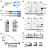
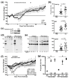
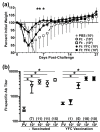
The targeting of immunogens/vaccines to specific immune cells is a promising approach for amplifying immune responses in the absence of exogenous adjuvants. However, the targeting approaches reported thus far require novel, labor-intensive reagents for each vaccine and have primarily been shown as proof-of-concept with isolated proteins and/or inactivated bacteria. We have engineered a plasmid-based, complement receptor-targeting platform that is readily applicable to live forms of multiple gram-negative bacteria, including, but not limited to, Escherichia coli, Klebsiella pneumoniae, and Francisella tularensis. Using F. tularensis as a model, we find that targeted bacteria show increased binding and uptake by macrophages, which coincides with increased p38 and p65 phosphorylation. Mice vaccinated with targeted bacteria produce higher titers of specific antibody that recognizes a greater diversity of bacterial antigens. Following challenge with homologous or heterologous isolates, these mice exhibited less weight loss and/or accelerated weight recovery as compared to counterparts vaccinated with non-targeted immunogens. Collectively, these findings provide proof-of-concept for plasmid-based, complement receptor-targeting of live gram-negative bacteria.
Keywords: autotransporter; complement; gram-negative; play; plug & tularemia; vaccine-targeting.
Conflict of interest statement
K.M.H.-T., E.J.G., and K.ROH. are inventors on a provisional patent for plasmids described in this manuscript. K.M.H.-T., L.E.S., D.S., S.K., S.J.R., P.N., A.S., T.J.S., E.J.G., and K.ROH. declare no conflict of interest.
Figures


Similar articles
-
Inactivated Francisella tularensis live vaccine strain protects against respiratory tularemia by intranasal vaccination in an immunoglobulin A-dependent fashion.Infect Immun. 2007 May;75(5):2152-62. doi: 10.1128/IAI.01606-06. Epub 2007 Feb 12. Infect Immun. 2007. PMID: 17296747 Free PMC article.
-
Live Attenuated Tularemia Vaccines for Protection Against Respiratory Challenge With Virulent F. tularensis subsp. tularensis.Front Cell Infect Microbiol. 2018 May 15;8:154. doi: 10.3389/fcimb.2018.00154. eCollection 2018. Front Cell Infect Microbiol. 2018. PMID: 29868510 Free PMC article. Review.
-
Aim2 and Nlrp3 Are Dispensable for Vaccine-Induced Immunity against Francisella tularensis Live Vaccine Strain.Infect Immun. 2021 Jun 16;89(7):e0013421. doi: 10.1128/IAI.00134-21. Epub 2021 Jun 16. Infect Immun. 2021. PMID: 33875472 Free PMC article.
-
Identification of Francisella tularensis outer membrane protein A (FopA) as a protective antigen for tularemia.Vaccine. 2011 Sep 16;29(40):6941-7. doi: 10.1016/j.vaccine.2011.07.075. Epub 2011 Jul 29. Vaccine. 2011. PMID: 21803089 Free PMC article.
-
Development of a Multivalent Subunit Vaccine against Tularemia Using Tobacco Mosaic Virus (TMV) Based Delivery System.PLoS One. 2015 Jun 22;10(6):e0130858. doi: 10.1371/journal.pone.0130858. eCollection 2015. PLoS One. 2015. PMID: 26098553 Free PMC article.
Cited by
-
The O-Ag Antibody Response to Francisella Is Distinct in Rodents and Higher Animals and Can Serve as a Correlate of Protection.Pathogens. 2021 Dec 20;10(12):1646. doi: 10.3390/pathogens10121646. Pathogens. 2021. PMID: 34959601 Free PMC article.
-
Co-Opting Host Receptors for Targeted Delivery of Bioconjugates-From Drugs to Bugs.Molecules. 2021 Mar 9;26(5):1479. doi: 10.3390/molecules26051479. Molecules. 2021. PMID: 33803208 Free PMC article. Review.
-
Francisella and Antibodies.Microorganisms. 2021 Oct 12;9(10):2136. doi: 10.3390/microorganisms9102136. Microorganisms. 2021. PMID: 34683457 Free PMC article. Review.
-
Current vaccine strategies and novel approaches to combatting Francisella infection.Vaccine. 2024 Apr 2;42(9):2171-2180. doi: 10.1016/j.vaccine.2024.02.086. Epub 2024 Mar 8. Vaccine. 2024. PMID: 38461051 Free PMC article. Review.
References
Grants and funding
LinkOut - more resources
Full Text Sources
Medical
Molecular Biology Databases

