. 2020 May 14;10(5):942.
doi: 10.3390/nano10050942.

Gold Nanorod Assemblies: The Roles of Hot-Spot Positioning and Anisotropy in Plasmon Coupling and SERS

Affiliations

Gold Nanorod Assemblies: The Roles of Hot-Spot Positioning and Anisotropy in Plasmon Coupling and SERS

Priyanka Dey et al. Nanomaterials (Basel). .

Abstract

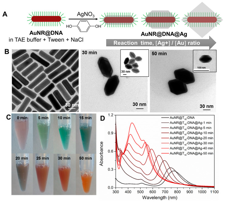
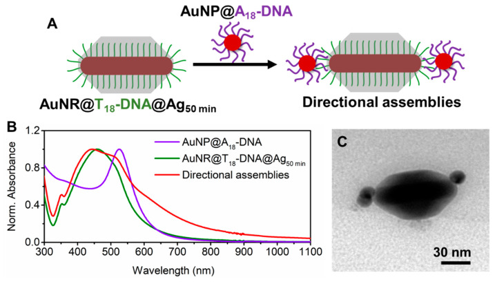
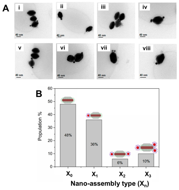
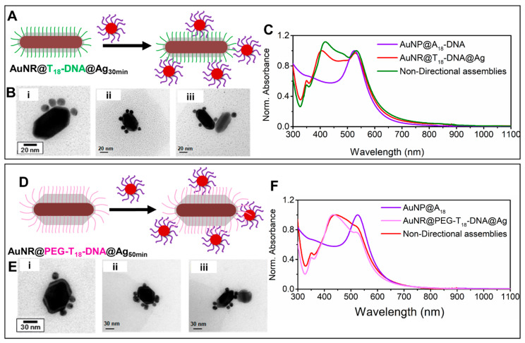
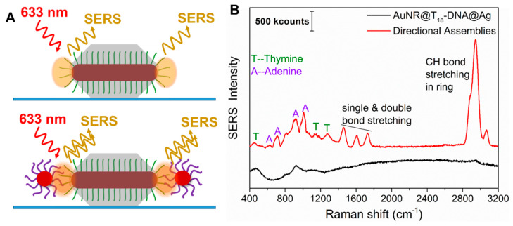
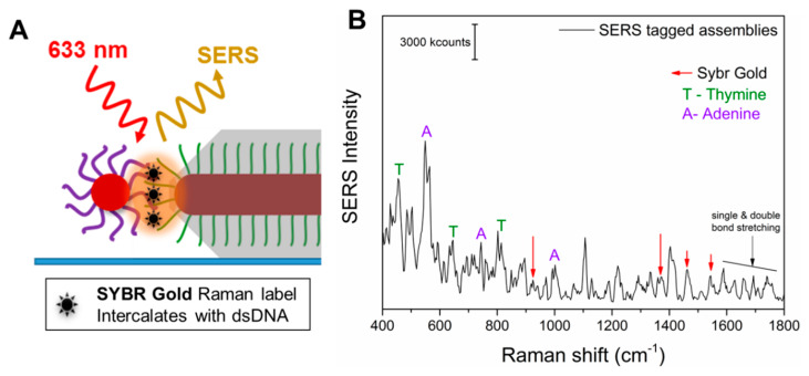
Plasmon-coupled colloidal nanoassemblies with carefully sculpted "hot-spots" and intense surface-enhanced Raman scattering (SERS) are in high demand as photostable and sensitive plasmonic nano-, bio-, and chemosensors. When maximizing SERS signals, it is particularly challenging to control the hot-spot density, precisely position the hot-spots to intensify the plasmon coupling, and introduce the SERS molecule in those intense hot-spots. Here, we investigated the importance of these factors in nanoassemblies made of a gold nanorod (AuNR) core and spherical nanoparticle (AuNP) satellites with ssDNA oligomer linkers. Hot-spot positioning at the NR tips was made possible by selectively burying the ssDNA in the lateral facets via controlled Ag overgrowth while retaining their hybridization and assembly potential at the tips. This strategy, with slight alterations, allowed us to form nanoassemblies that only contained satellites at the NR tips, i.e., directional anisotropic nanoassemblies; or satellites randomly positioned around the NR, i.e., nondirectional nanoassemblies. Directional nanoassemblies featured strong plasmon coupling as compared to nondirectional ones, as a result of strategically placing the hot-spots at the most intense electric field position of the AuNR, i.e., retaining the inherent plasmon anisotropy. Furthermore, as the dsDNA was located in these anisotropic hot-spots, this allowed for the tag-free detection down to 10 dsDNA and a dramatic SERS enhancement of 1.6 × 10⁸ for the SERS tag SYBR gold, which specifically intercalates into the dsDNA. This dramatic SERS performance was made possible by manipulating the anisotropy of the nanoassemblies, which allowed us to emphasize the critical role of hot-spot positioning and SERS molecule positioning in nanoassemblies.

Keywords: DNA linker; SERS molecule positioning; anisotropy; core–satellite nanoassembly; directional assembly; gold nanorods; hot-spot positioning; silver coating.


Conflict of interest statement

The authors declare no conflict of interest.

Figures

Figure 1
Figure 1
(A) Sketch showing the gradual evolution of AuNRs@DNA into directional building blocks by epitaxial Ag overgrowth. By controlling the reaction time and Ag+/Au ratio, the resulting AuNRs@DNA@Ag will be: nondirectional (truncated octahedra with all DNA strands available for hybridization, left); directional (truncated octahedra with the DNA strands at the tip region available for hybridization, center); or partially directional, but colloidally unstable (nontruncated octahedra with most DNA strands inactivated for hybridization, right). (B) TEM micrographs depicting the starting AuNRs@T18-DNA (0 min, left panel) and the resulting AuNRs@T18-DNA@Ag at an intermediate reaction time (30 min) and after completion of the reaction (50 min). (C) Photographs illustrating the color changes of the AuNRs@T18-DNA dispersion (0 min) at the indicated reaction times. (D) Corresponding UV–Vis–NIR spectra during Ag overgrowth.
Figure 2
Figure 2
(A) Schematic illustration of the formation of anisotropic directional nanoassemblies upon incubation of AuNRs@T18-DNA@Ag (overgrown for 50 min) with AuNPs@A18-DNA. (B) UV–Vis–NIR spectra of each building block and the resulting directional anisotropic nanoassemblies. (C) Representative TEM micrograph of the obtained anisotropic directional nanoassemblies.
Figure 3
Figure 3
(A) (i–viii) Representative TEM micrographs of the anisotropic directional nanoassemblies obtained upon incubation of core AuNRs@T18-DNA@Ag (overgrown for 50 min) with satellite AuNPs@A18-DNA. (B) Nanoassembly yield (determined from statistical TEM analysis, population count = 100), resulting from the DNA hybridization of AuNRs@T18-DNA@Ag (overgrown for 50 min) with AuNPs@A18-DNA as a function of nanoassembly type Xn, where n = number of spherical satellite NPs per assembly. Overall, the anisotropic directional nanoassembly yield was ~52% (X1, X2, X3).
Figure 4
Figure 4
Nondirectional nanoassemblies obtained upon incubation of AuNRs@T18-DNA@Ag (overgrown for 30 min, left panel) or of AuNRs@PEG-T18-DNA@Ag (overgrown for 50 min, right panel) with AuNPs@A18-DNA. (A,D) Schematic illustration of the formation of nondirectional nanoassemblies for both cases. (B,E) Representative TEM micrographs of the corresponding nondirectional nanoassemblies. (C,F) UV–Vis–NIR spectra of the respective building blocks and their resulting nondirectional nanoassemblies.
Figure 5
Figure 5
(A) Sketch of the anisotropic directional building blocks (AuNRs@T18-DNA@Ag50min, top) and their anisotropic directional nanoassemblies (bottom). The orange shading highlights the spots of highest electric-field enhancement, i.e., the strategically positioned hot-spots that can lead to SERS of the ssDNA (top panel) or of the dsDNA (bottom panel). (B) Representative SERS spectra of a single AuNRs@T18-DNA@Ag (directional building block, black curve) and of a single anisotropic directional nanoassembly (red curve), as investigated by dark-field microscope (DFM)-SERS (λlaser = 633 nm). The characteristic SERS peaks from A and T are indicated.
Figure 6
Figure 6
(A) Magnified depiction of a SYBR-gold-tagged anisotropic directional nanoassembly spin-coated onto a glass substrate. The SYBR gold intercalates within the dsDNA. The plasmonic hot-spot is depicted in orange. (B) Representative SERS spectrum from a SYBR-gold-tagged directional nanoassembly. The signature peaks of adenine (A), thymine (T), and SYBR gold are indicated.

References

    1. Vo-dinh T., Liu Y., Crawford B.M., Wang H., Yuan H., Register J.K., Zhang Y., Liu Z.Z., Thackray B.D., Bao Z., et al. Rapid, One-Pot, Protein-Mediated Green Synthesis of Gold Nanostars for Computed Tomographic Imaging and Photothermal Therapy of Cancer. Chem. Soc. Rev. 2017;44:386–406.
    1. Guerrini L., Graham D. Molecularly-Mediated Assemblies of Plasmonic Nanoparticles for Surface-Enhanced Raman Spectroscopy Applications. Chem. Soc. Rev. 2012;41:7085–7107. doi: 10.1039/c2cs35118h. - DOI - PubMed
    1. Sajanlal P.R., Sreeprasad T.S., Samal A.K., Pradeep T. Anisotropic Nanomaterials: Structure, Growth, Assembly, and Functions. Nano Rev. 2011;2:5883. doi: 10.3402/nano.v2i0.5883. - DOI - PMC - PubMed
    1. Pansare V., Hejazi S., Faenza W., Prud’homme R.K. Review of Long-Wavelength Optical and NIR Imaging Materials: Contrast Agents, Fluorophores and Multifunctional Nano Carriers. Chem. Mater. 2012;24:812–827. doi: 10.1021/cm2028367. - DOI - PMC - PubMed
    1. Dey P., Tabish T.A., Mosca S., Palombo F., Matousek P., Stone N. Plasmonic Nanoassemblies: Tentacles Beat Satellites for Boosting Broadband NIR Plasmon Coupling Providing a Novel Candidate for SERS and Photothermal Therapy. Small. 2020;16:1906780. doi: 10.1002/smll.201906780. - DOI - PubMed

LinkOut - more resources