Review
. 2020 Jul 3;31(5):580-588.
doi: 10.1080/09537104.2020.1763939. Epub 2020 May 18.

Use of electron microscopy to study platelets and thrombi

Affiliations
Review

Use of electron microscopy to study platelets and thrombi

Maurizio Tomaiuolo et al. Platelets. .

Abstract

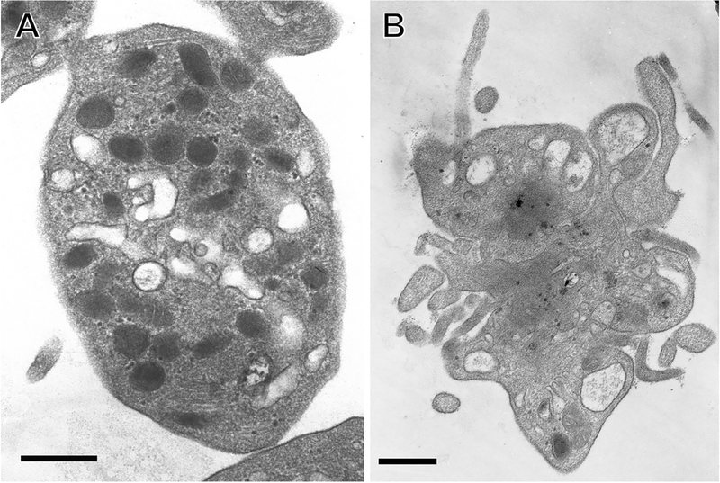
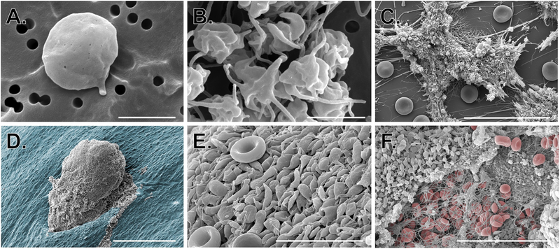
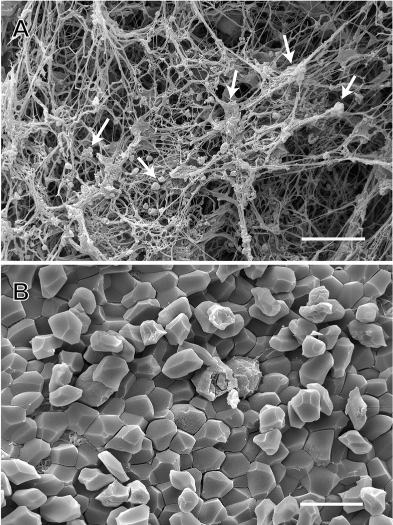
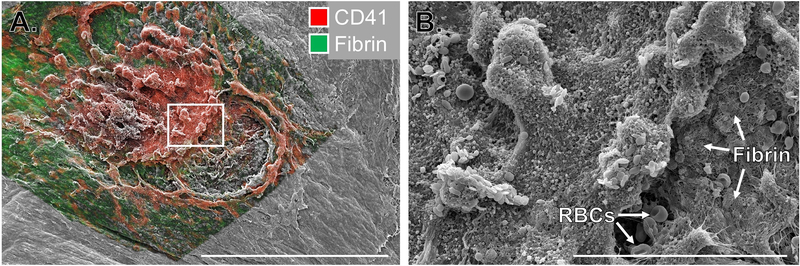
Electron microscopy has been a valuable tool for the study of platelet biology and thrombosis for more than 70 years. Early studies using conventional transmission and scanning electron microscopy (EM) provided a foundation for our initial understanding of platelet structure and how it changes upon platelet activation. EM approaches have since been utilized to study platelets and thrombi in the context of basic, translational and clinical research, and they are instrumental in the diagnosis of multiple platelet function disorders. In this brief review, we provide a sampling of the many contributions EM based studies have made to the field, including both historical highlights and contemporary applications. We will also discuss exciting new imaging modalities based on EM and their utility for the study of platelets, hemostasis and thrombosis into the future.

Keywords: Hemostasis; platelet; scanning electron microscopy; thrombosis; transmission electron microscopy.


Figures

Figure 1:
Figure 1:. Use of TEM to study platelet structure.
TEM images show A) isolated resting human platelet; B) isolated thrombin-activated human platelet. Scale bars = 500 nm. Image credit: from the laboratory of John W. Weisel: Anastasia A. Ponomareva and Rustem I. Litvinov.
Figure 2:
Figure 2:. Use of SEM to study platelets in vitro and in vivo.
SEM images show A-B) isolated resting and activated human platelets (scale bars = 2 μm); C) human platelet-rich thrombi formed in a microfluidic flow chamber coated with collagen and tissue factor (scale bar = 30 μm); D-F) platelet-rich hemostatic plugs formed following puncture injury to a mouse jugular vein. D) hemostatic plug imaged from the intraluminal side (scale bar = 150 μm); the endothelium is pseudocolored blue. E) High magnification image of platelets on the luminal surface of a hemostatic plug (red blood cells can be seen for comparison; scale bar = 10 μm). F) Extraluminal component of a hemostatic plug showing platelets, fibrin and a few fibrin entrapped red blood cells (RBCs are pseudocolored red, scale bar = 30 μm). Image credit: M. Tomaiuolo and T.J. Stalker.
Figure 3:
Figure 3:. SEM of arterial thrombi taken from patients by thrombectomy.
SEM images show A) single and aggregated platelets (white arrows) and fibrin in an arterial thrombus; an example of the appearance of the outside of a contracted blood clot or thrombus; B) polyhedrocytes (compressed polyhedral erythrocytes) in an arterial thrombus; an example of the interior of a contracted blood clot. Scale bars = 10 μm. Image credit: from the laboratory of John W. Weisel: Rafael R. Khismatullin and Rustem I. Litvinov.
Figure 4:
Figure 4:. Correlative light and electron microscopy to study hemostatic plug architecture.
Images show the extravascular surface of a mouse jugular vein following puncture injury. A) Multiphoton fluorescence image of platelets (CD41, red) and fibrin (green) overlaid on an SEM image of the same region. Scale bar is 300 μm. B) SEM image of the region indicated by the white rectangle in A. Scale bar is 50 μm. Images adapted from Tomaiuolo et al [52].

Similar articles

Cited by

References

    1. Bessis M *Ultra-Structure Du Protoplasma Des Thrombocytes Au Microscope Electronique. Comptes Rendus Des Seances De La Societe De Biologie Et De Ses Filiales. 1948;142(9–10):647–649. - PubMed
    1. White JG, Krumwiede M. Some contributions of electron microscopy to knowledge of human platelets. Thromb Haemost. 2007;98(1):69–72. - PubMed
    1. White JG. Use of the electron microscope for diagnosis of platelet disorders. Semin Thromb Hemost. 1998;24(2):163–8. - PubMed
    1. Scandola C, Erhardt M, Rinckel JY, Proamer F, Gachet C, Eckly A. Use of electron microscopy to study megakaryocytes. Platelets. 2020:1–10. - PubMed
    1. Bessis M Studies in electron microscopy of blood cells. Blood. 1950;5(12):1083–98. - PubMed

MeSH terms