

Aggressiveness Potential of Spontaneous Canine Mucosal Melanoma Can Dictate Distinct Cancer Stem Cell Compartment Behaviors in Regard to Their Initial Size and Expansion Abilities
- PMID: 32423311
- PMCID: PMC7374591
- DOI: 10.1089/scd.2019.0223
Aggressiveness Potential of Spontaneous Canine Mucosal Melanoma Can Dictate Distinct Cancer Stem Cell Compartment Behaviors in Regard to Their Initial Size and Expansion Abilities
Abstract
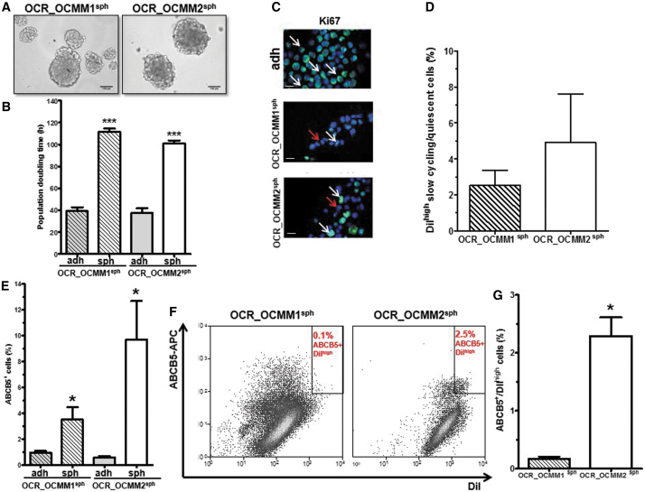
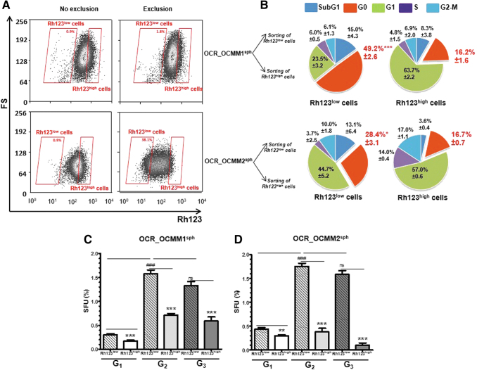
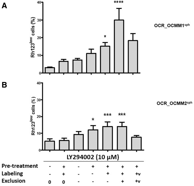
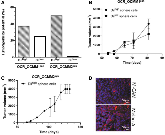
Mucosal melanoma represents one of the most highly metastatic and aggressive subtypes of melanoma. The biology of mucosal melanoma is poorly documented, and the lack of experimental models makes it difficult to design and test new therapies. Dogs are frequently affected by melanomas of the oral cavity, making spontaneous canine melanoma a potentially predictable model for their human counterpart. We recently established and characterized two new canine mucosal melanoma cell lines named OCR_OCMM1 and OCR_OCMM2. Here, we identified quiescent cancer stem cell (CSC) subpopulations in both canine cell lines that displayed similarities to human quiescent CSCs: canine melanoma CSCs had the ability to self-renew, produced nonstem cell (SC) progeny, and formed melanospheres that recapitulated the phenotypic profile of the parental tumor. These CSCs also formed melanoma in immunodeficient mice, and the inhibition of PI3K/AKT signaling expanded the CSC pool. A subset of non-CSCs transitioned to become CSCs. OCR_OCMM1 and OCR_OCMM2 displayed different CSC compartment behaviors in regard to their initial size and expansion abilities. Collectively, this study showed that the OCR_OCMM1 and OCR_OCMM2 canine melanoma cell lines are powerful cellular tools to study melanoma SCs, not only for mucosal but also for the more common human cutaneous melanoma.
Keywords: comparative oncology; melanoma; stem cells.
Conflict of interest statement
No competing financial interests exist.
Figures
Similar articles
-
The PI3K/AKT signaling pathway controls the quiescence of the low-Rhodamine123-retention cell compartment enriched for melanoma stem cell activity.Stem Cells. 2013 Apr;31(4):641-51. doi: 10.1002/stem.1333. Stem Cells. 2013. PMID: 23355370
-
Isolation and characterization of two canine melanoma cell lines: new models for comparative oncology.BMC Cancer. 2018 Dec 4;18(1):1219. doi: 10.1186/s12885-018-5114-y. BMC Cancer. 2018. PMID: 30514258 Free PMC article.
-
Synergistic targeted inhibition of MEK and dual PI3K/mTOR diminishes viability and inhibits tumor growth of canine melanoma underscoring its utility as a preclinical model for human mucosal melanoma.Pigment Cell Melanoma Res. 2016 Nov;29(6):643-655. doi: 10.1111/pcmr.12512. Epub 2016 Sep 22. Pigment Cell Melanoma Res. 2016. PMID: 27463366 Free PMC article.
-
Targeting the phosphatidylinositol 3-kinase/Akt/mammalian target of rapamycin signaling network in cancer stem cells.Curr Med Chem. 2011;18(18):2715-26. doi: 10.2174/092986711796011201. Curr Med Chem. 2011. PMID: 21649579 Review.
-
Cellular and molecular biology of cancer stem cells in melanoma: Possible therapeutic implications.Semin Cancer Biol. 2019 Dec;59:221-235. doi: 10.1016/j.semcancer.2019.06.019. Epub 2019 Jun 29. Semin Cancer Biol. 2019. PMID: 31265892 Review.
Cited by
-
Involvement of ORAI1/SOCE in Human AML Cell Lines and Primary Cells According to ABCB1 Activity, LSC Compartment and Potential Resistance to Ara-C Exposure.Int J Mol Sci. 2022 May 16;23(10):5555. doi: 10.3390/ijms23105555. Int J Mol Sci. 2022. PMID: 35628366 Free PMC article.
-
Specific oncogene activation of the cell of origin in mucosal melanoma.bioRxiv [Preprint]. 2024 Apr 26:2024.04.22.590595. doi: 10.1101/2024.04.22.590595. bioRxiv. 2024. Update in: Nat Commun. 2025 Jul 22;16(1):6750. doi: 10.1038/s41467-025-61937-1. PMID: 38712250 Free PMC article. Updated. Preprint.
-
Characterization of Primary Cultures of Normal and Neoplastic Canine Melanocytes.Animals (Basel). 2021 Mar 10;11(3):768. doi: 10.3390/ani11030768. Animals (Basel). 2021. PMID: 33802040 Free PMC article.
-
Novel cellular systems unveil mucosal melanoma initiating cells and a role for PI3K/Akt/mTOR pathway in mucosal melanoma fitness.J Transl Med. 2024 Jan 8;22(1):35. doi: 10.1186/s12967-023-04784-2. J Transl Med. 2024. PMID: 38191367 Free PMC article.
-
A Comparative View on Molecular Alterations and Potential Therapeutic Strategies for Canine Oral Melanoma.Vet Sci. 2021 Nov 22;8(11):286. doi: 10.3390/vetsci8110286. Vet Sci. 2021. PMID: 34822659 Free PMC article. Review.
References
-
- Reya T, Morrison SJ, Clarke MF and Weissman IL (2001). Stem cells, cancer, and cancer stem cells. Nature 414:105–111 - PubMed
-
- Hoek KS and Goding CR (2010). Cancer stem cells versus phenotype-switching in melanoma. Pigment Cell Melanoma Res 23:746–759 - PubMed
-
- Clevers H. (2011). The cancer stem cell: premises, promises and challenges. Nat Med 17:313–319 - PubMed
-
- Touil Y, Zuliani T, Wolowczuk I, Kuranda K, Prochazkova J, Andrieux J, Le Roy H, Mortier L, Vandomme J, et al. (2013). The PI3K/AKT signaling pathway controls the quiescence of the low-Rhodamine123-retention cell compartment enriched for melanoma stem cell activity. Stem Cells 31:641–651 - PubMed
Publication types
MeSH terms
Substances
LinkOut - more resources
Full Text Sources
Medical