Review
. 2020 Sep;40(9):1752-1768.
doi: 10.1177/0271678X20923551. Epub 2020 May 19.

Potential therapeutic targets for intracerebral hemorrhage-associated inflammation: An update

Affiliations
Review

Potential therapeutic targets for intracerebral hemorrhage-associated inflammation: An update

Honglei Ren et al. J Cereb Blood Flow Metab. 2020 Sep.

Abstract

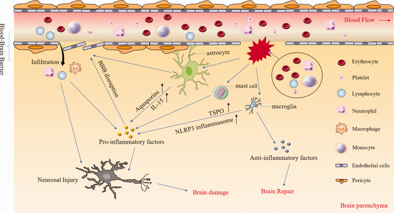
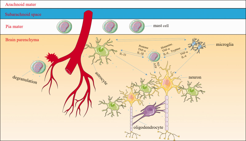
Intracerebral hemorrhage (ICH) is a subtype of stroke with high mortality and disability but no specific or effective treatment. In the last two decades, much has been learned about the pathologic mechanisms of ICH. It is now known that after ICH onset, immune and inflammatory responses contribute to blood-brain barrier disruption, edema development, and cell death processes, jointly resulting in secondary brain injury. However, the translation of potential therapies from preclinical to clinical success has been disappointing. With the development of new laboratory technology, recent progress has been made in the understanding of ICH pathomechanisms, and promising therapeutic targets have been identified. This review provides an update of recent progress on ICH and describes the prospects for further preclinical studies in this field. Our goal is to discuss new therapeutic targets and directions for the treatment of ICH and promote the effective transformation from preclinical to clinical trials.

Keywords: Intracerebral hemorrhage; immune interventions; inflammation; microglia; secondary injury.


Figures

Figure 1.
Figure 1.
Inflammatory responses after intracerebral hemorrhage (ICH). After ICH, multiple blood components (e.g. erythrocytes, leukocytes, platelets) are released into the brain parenchyma where they can activate microglia, astrocytes, and mast cells by distinct pathways. After activation, microglia upregulate the expression of NLRP3 and TSPO and produce large amounts of pro-inflammatory factors. However, they also produce anti-inflammatory factors such as TGF-β which contributes to brain repair. Astrocytes contribute to the composition of the blood–brain barrier (BBB) and are destroyed directly after ICH, leading to BBB disruption. In addition, astrocytes secrete various pro-inflammatory factors that aggravate neuronal injury and brain damage. Pro-inflammatory factors from diverse cell types not only exacerbate BBB disruption and leukocyte infiltration, but also kill neurons directly. NLRP3: the nucleotide-binding oligomerization domain-like receptor containing pyrin domain 3; TSPO: translocator protein.
Figure 2.
Figure 2.
Mast cells (MCs) might be some of the central mediators of inflammatory responses. MCs are normal resident cells in the leptomeninges and brain structures surrounding blood vessels. After the onset of intracerebral hemorrhage (ICH), MCs rapidly respond by degranulating and releasing preformed and stored mediators including histamine, serotonin, heparin, TNF-α, and other cytokines. MC-derived granules can activate both microglia and astrocytes as well as exacerbate neuronal necrosis and apoptosis. For example, MC-derived tryptase induces microglial activation and secretion of pro-inflammatory factors via the protease-activated receptor 2 signaling pathway; MC-derived histamine stimulates microglial activation and the subsequent production of pro-inflammatory factors. MC proteases activate astrocytes and cause the release of IL-33 by activating p38, ERK1/2 MAPKs, and the NF-κB signaling pathway. In turn, activated microglia and astrocytes further stimulate MCs to release histamine, IL-6, and TNF-α. The interaction between MCs and oligodendrocytes is still unclear. The MC–microglia–astrocyte network amplifies the cascade of inflammatory responses and aggravates neuronal necrosis and apoptosis after ICH.

References

    1. Keep RF, Hua Y, Xi G.Intracerebral haemorrhage: mechanisms of injury and therapeutic targets. Lancet Neurol 2012; 11: 720–731. - PMC - PubMed
    1. Wang J.Preclinical and clinical research on inflammation after intracerebral hemorrhage. Prog Neurobiol 2010; 92: 463–477. - PMC - PubMed
    1. Zhou Y, Wang Y, Wang J, et al.. Inflammation in intracerebral hemorrhage: from mechanisms to clinical translation. Prog Neurobiol 2014; 115: 25–44. - PubMed
    1. Jolink WM, Klijn CJ, Brouwers PJ, et al.. Time trends in incidence, case fatality, and mortality of intracerebral hemorrhage. Neurology 2015; 85: 1318–1324. - PubMed
    1. Marini S, Morotti A, Ayres AM, et al.. Sex differences in intracerebral hemorrhage expansion and mortality. J Neurol Sci 2017; 379: 112–116. - PMC - PubMed

Publication types