

Chest CT imaging characteristics of COVID-19 pneumonia in preschool children: a retrospective study
- PMID: 32423435
- PMCID: PMC7232932
- DOI: 10.1186/s12887-020-02140-7
Chest CT imaging characteristics of COVID-19 pneumonia in preschool children: a retrospective study
Abstract
Background: Recently, the World Health Organization has declared the coronavirus disease 2019 (COVID-19) outbreak a public health emergency of international concern. So far, however, limited data are available for children. Therefore, we aimed to investigate the clinical and chest CT imaging characteristics of COVID-19 in preschool children.
Methods: From January 26, 2020 to February 20, 2020, the clinical and initial chest CT imaging data of eight preschool children with laboratory-confirmed COVID-19 from two hospitals were retrospectively collected. The chest CT imaging characteristics, including the distribution, shape, and density of lesions, and the pleural effusion, pleural changes, and enlarged lymph nodes were evaluated.
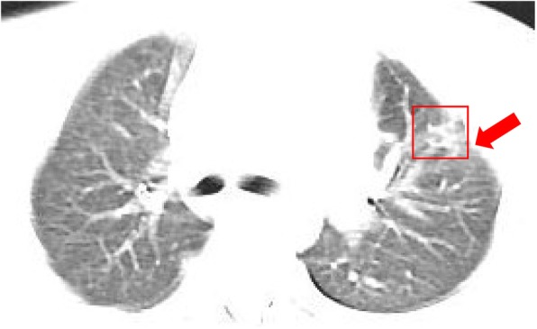
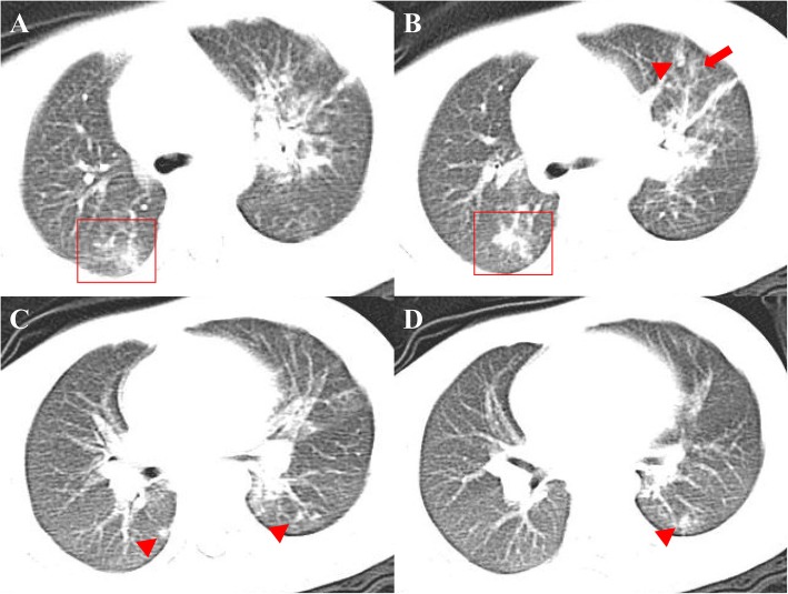
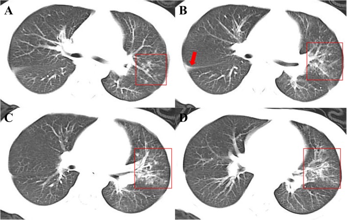
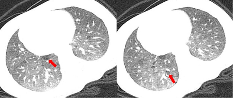
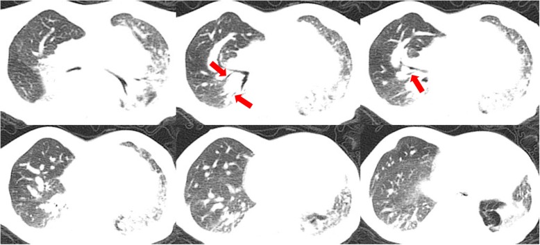
Results: Two cases (25%) were classified as mild type, and they showed no obvious abnormal CT findings or minimal pleural thickening on the right side. Five cases (62.5%) were classified as moderate type. Among these patients, one case showed consolidation located in the subpleural region of the right upper lobe, with thickening in the adjacent pleura; one case showed multiple consolidation and ground-glass opacities with blurry margins; one case displayed bronchial pneumonia-like changes in the left upper lobe; and two cases displayed asthmatic bronchitis-like changes. One case (12.5%) was classified as critical type and showed bronchial pneumonia-like changes in the bilateral lungs, presenting blurred and messy bilateral lung markings and multiple patchy shadows scattered along the lung markings with blurry margins.
Conclusions: The chest CT findings of COVID-19 in preschool children are atypical and various. Accurate diagnosis requires a comprehensive evaluation of epidemiological, clinical, laboratory and CT imaging data.
Keywords: COVID-19; Child; Multidetector computed tomography; Pneumonia; SARS-CoV-2.
Conflict of interest statement
The authors declare that they have no competing interests.
Figures
Similar articles
-
Imaging and clinical features of patients with 2019 novel coronavirus SARS-CoV-2.Eur J Nucl Med Mol Imaging. 2020 May;47(5):1275-1280. doi: 10.1007/s00259-020-04735-9. Epub 2020 Feb 28. Eur J Nucl Med Mol Imaging. 2020. PMID: 32107577 Free PMC article.
-
Clinical and computed tomographic imaging features of novel coronavirus pneumonia caused by SARS-CoV-2.J Infect. 2020 Apr;80(4):394-400. doi: 10.1016/j.jinf.2020.02.017. Epub 2020 Feb 25. J Infect. 2020. PMID: 32109443 Free PMC article.
-
[CT imaging analysis of 33 cases with the 2019 novel coronavirus infection].Zhonghua Yi Xue Za Zhi. 2020 Apr 7;100(13):1007-1011. doi: 10.3760/cma.j.cn112137-20200203-00182. Zhonghua Yi Xue Za Zhi. 2020. PMID: 32294858 Chinese.
-
Thoracic imaging tests for the diagnosis of COVID-19.Cochrane Database Syst Rev. 2020 Sep 30;9:CD013639. doi: 10.1002/14651858.CD013639.pub2. Cochrane Database Syst Rev. 2020. Update in: Cochrane Database Syst Rev. 2020 Nov 26;11:CD013639. doi: 10.1002/14651858.CD013639.pub3. PMID: 32997361 Updated.
-
Chest computed tomography findings in hospitalized COVID-19 patients: a systematic review and meta-analysis.Infez Med. 2020 Sep 1;28(3):295-301. Infez Med. 2020. PMID: 32920564
Cited by
-
Radiological Findings of COVID-19 in Children: A Systematic Review and Meta-Analysis.J Trop Pediatr. 2021 Jul 2;67(3):fmaa045. doi: 10.1093/tropej/fmaa045. J Trop Pediatr. 2021. PMID: 32692815 Free PMC article.
-
A Single-Center (Sibiu, Romania), Retrospective Study (March-November 2020) of COVID-19 Clinical and Epidemiological Features in Children.J Clin Med. 2021 Aug 10;10(16):3517. doi: 10.3390/jcm10163517. J Clin Med. 2021. PMID: 34441813 Free PMC article.
-
Pediatric lung imaging features of COVID-19: A systematic review and meta-analysis.Pediatr Pulmonol. 2021 Jan;56(1):252-263. doi: 10.1002/ppul.25070. Epub 2020 Nov 2. Pediatr Pulmonol. 2021. PMID: 32926572 Free PMC article.
-
Imaging findings in acute pediatric coronavirus disease 2019 (COVID-19) pneumonia and multisystem inflammatory syndrome in children (MIS-C).Pediatr Radiol. 2022 Sep;52(10):1985-1997. doi: 10.1007/s00247-022-05393-9. Epub 2022 May 26. Pediatr Radiol. 2022. PMID: 35616701 Free PMC article. Review.
-
Chest computed tomography (CT) features in children with reverse transcription-polymerase chain reaction (RT-PCR)-confirmed COVID-19: A systematic review.J Med Imaging Radiat Oncol. 2020 Oct;64(5):649-659. doi: 10.1111/1754-9485.13098. Epub 2020 Sep 30. J Med Imaging Radiat Oncol. 2020. PMID: 33000560 Free PMC article.
References
-
- World Health Organization: Coronavirus disease (COVID-19) outbreak. 2020, (https://www.who.int).
-
- Li Y, Xia L. Coronavirus Disease 2019 (COVID-19): role of chest CT in diagnosis and management. Am J Roentgenol. 2020(4):1–7. - PubMed
MeSH terms
LinkOut - more resources
Full Text Sources
Medical
Miscellaneous
