Review
. 2020 Jul;43(7):458-466.
doi: 10.1016/j.tins.2020.04.003. Epub 2020 May 15.

The Brain's Glymphatic System: Current Controversies

Affiliations
Review

The Brain's Glymphatic System: Current Controversies

Humberto Mestre et al. Trends Neurosci. 2020 Jul.

Abstract

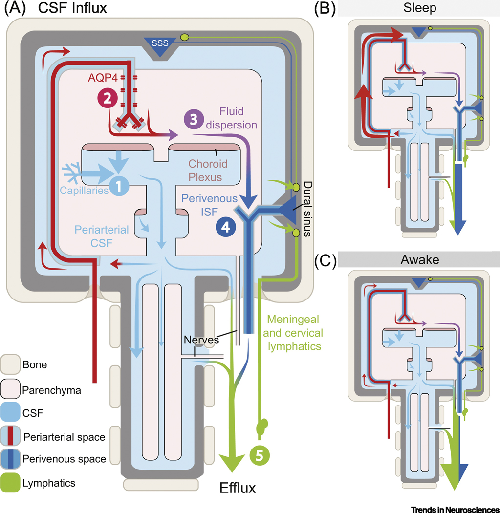
The glymphatic concept along with the discovery of meningeal lymphatic vessels have, in recent years, highlighted that fluid is directionally transported within the central nervous system (CNS). Imaging studies, as well as manipulations of fluid transport, point to a key role of the glymphatic-lymphatic system in clearance of amyloid-β and other proteins. As such, the glymphatic-lymphatic system represents a new target in combating neurodegenerative diseases. Not unexpectedly, introduction of a new plumbing system in the brain has stirred controversies. This opinion article will highlight what we know about the brain's fluid transport systems, where experimental data are lacking, and what is still debated.

Keywords: amyloid-β; aquaporin-4; brain clearance; cerebrospinal fluid; glymphatic system; meningeal lymphatics; perivascular spaces.


Conflict of interest statement

Conflicts of interest

The authors have no conflicts of interest to report.

Figures

Fig. 1.
Fig. 1.. Schematic model of the glymphatic-lymphatic system and its regulation by the sleep-wake cycle.
(A) The fluid transport pathway is divided into 5 distinct segments: (1) cerebrospinal fluid (CSF) is produced by the choroid plexus and likely by extrachoroidal sources (capillary influx and metabolic water production); (2) arterial wall pulsatility drives CSF deep into brain along perivascular spaces; (3) CSF enters the brain parenchyma supported by AQP4 water channels and disperses within the neuropil; (4) interstitial fluid (ISF) mixes with CSF and accumulates in the peri-venous space and drains from here out of the brain via (5) meningeal and cervical lymphatic vessels, as well as along cranial and spinal nerves. The two models on the right, map the dominant paths of CSF flow during different arousal states: during sleep (B), CSF enters the brain via glymphatic transport, but during wakefulness (C) it is mostly excluded and shunted out via lymphatic vessels [4, 43]. The exact fraction of CSF entering brain during sleep is not known, but MRI studies suggest that ~20% of contrast agents injected into cisterna magna are taken up by the brain in anesthetized rats [62]. Recent evidence collected in awake rats using contrast-enhanced MRI shows that the glymphatic system is also under circadian control [63]. According to an older, but debated literature, CSF can exit via arachnoid granulations not included in this model [64, 65]. AQP4: Aquaporin-4 channels; CSF: cerebrospinal fluid; ISF: interstitial fluid; SSS: superior sagittal sinus.
Fig. 2.
Fig. 2.. Diagram illustrating postmortem relocation of periarterial cerebrospinal fluid (CSF) tracers.
In the live brain (left), the pulsatility of pial arteries drives CSF tracers along large surface perivascular spaces (A–C; B from [66], A, C from [7]). The tracers are confined to the perivascular spaces on each side of the vessel and do not enter the compact smooth muscle cell layer (C inset, orthogonal projection). CSF tracers enter the neuropil by leaving the perivascular space (PVS) of penetrating arteries via gaps between the vascular endfeet of astrocytes (D–F; panels D, E, F are from [19], [20], and [18], respectively). Postmortem (right), the arteries collapse, and the fluid filled PVS largely disappears (G–I; compare to A–C. G, I from [7]; H from [66]), and tracers relocate into the smooth muscle cell (SMC) layer, basal lamina, and endothelial cell (EC) basement membrane (J–L; panels J, K and L are from [1], [67], and [68], respectively). Please refer to original publications for further details.
Fig. 3.
Fig. 3.. Intracranial pressure (ICP) is highly dynamic in live rodents.
ICP pulsates in synchrony with both the cardiac and the respiratory cycle (Δ2 mmHg)[69]. (A) ICP undergoes a dramatic 3-fold increase during development, increasing in mice from 1.33±0.87 mmHg at postnatal day 3 (P3) to 4.11±0.83 mmHg at P70. Data adapted from Fig. 2 in[70]. (B) ICP is highly dependent on postural changes in rats anesthetized with isoflurane. A −45° head tilt down can elevate ICP by 4 mmHg while a +45° head tilt up can likewise decrease it 4 mmHg (time scale: 7.8 and 10 min, respectively). Adapted from Fig. 2b in[69]. (C, left panel) Respiratory acidosis or hypercapnia (high pCO2) causes cerebral blood vessels to dilate, increasing intracranial volume and in turn ICP. Hypercapnia induced by inhaling 5% CO2 in air causes a ~15 mmHg increase in baseline ICP. Right panel, chest compressions or Valsalva maneuvers increase intrathoracic pressure and reduce cerebral venous outflow elevating ICP ~9 mmHg. Adapted from Fig. 1d in [71]. (D) Top: dynamic-contrast enhanced MRI after gadobutrol intracisternal injection (12 μl at 1.5μl/min) shown as an enhancement ratio[5]. Bottom: ICP during infusion of 10 μl of tracer into cisterna magna at 1 μl/min or 2 μl/min. Intracisternal injection increases ICP by 1.4–3 mmHg and returns to baseline within 5 min after the end of the infusion. N= 5–6 mice per group. CSF influx takes place up to 20 min after the end of the ICP increase. (E) Summary of mean variations in ICP (mmHg) in response to different manipulations or state changes: respiration (Resp), development (Dev), postural changes as in panel (b), hypercapnia and Valsalva maneuvers as in panel (c), and tracer infusions as in (d). Changes in ICP caused by tracer infusion are relatively modest, and the overall comparison suggests that commonplace physiological changes for example in body posture or arterial blood gases are stronger modifiers of the glymphatic/lymphatic fluid transport than tracer injection.

Similar articles

Cited by

References

    1. Iliff JJ et al. (2012) A paravascular pathway facilitates CSF flow through the brain parenchyma and the clearance of interstitial solutes, including amyloid beta. Sci Transl Med 4 (147), 147ra111. - PMC - PubMed
    1. Louveau A et al. (2015) Structural and functional features of central nervous system lymphatic vessels. Nature 523 (7560), 337–41. - PMC - PubMed
    1. Aspelund A et al. (2015) A dural lymphatic vascular system that drains brain interstitial fluid and macromolecules. J Exp Med 212 (7), 991–9. - PMC - PubMed
    1. Ma Q et al. (2019) Rapid lymphatic efflux limits cerebrospinal fluid flow to the brain. Acta Neuropathol 137 (1), 151–165. - PMC - PubMed
    1. Mestre H et al. (2020) Cerebrospinal fluid influx drives acute ischemic tissue swelling. Science 367 (6483). - PMC - PubMed

Publication types