Case Reports
. 2020 Aug;26(3):e189-e194.
doi: 10.1016/j.radi.2020.04.017. Epub 2020 May 11.

False negative chest X-Rays in patients affected by COVID-19 pneumonia and corresponding chest CT findings

Affiliations
Case Reports

False negative chest X-Rays in patients affected by COVID-19 pneumonia and corresponding chest CT findings

M Cellina et al. Radiography (Lond). 2020 Aug.

Abstract

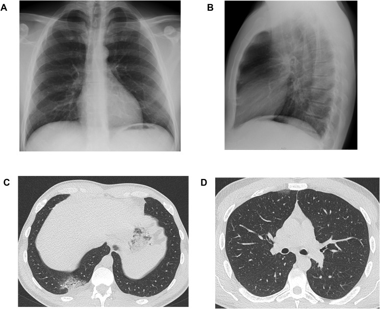
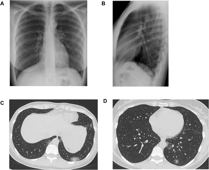
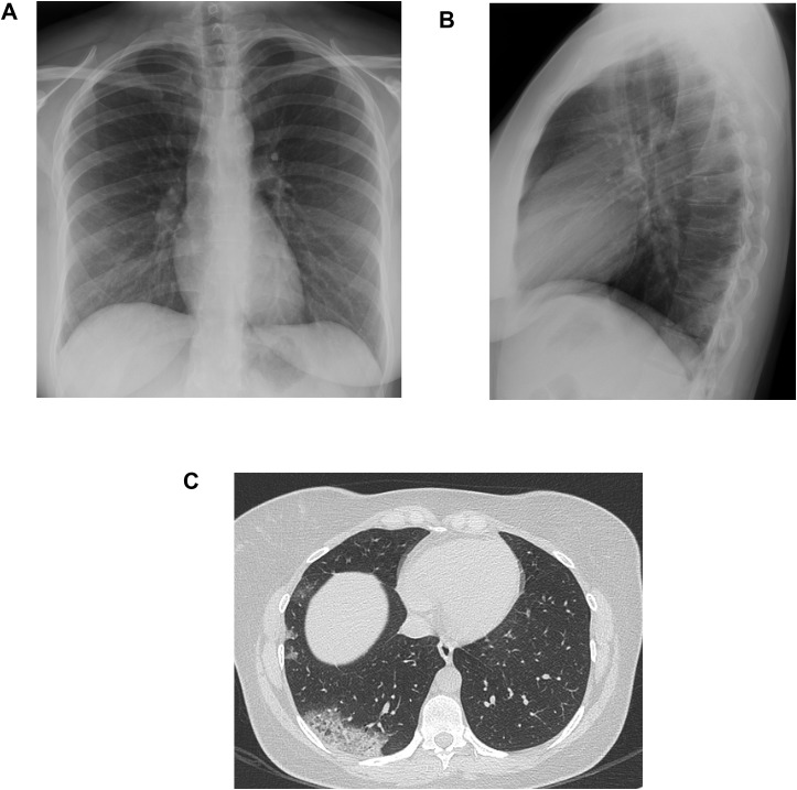
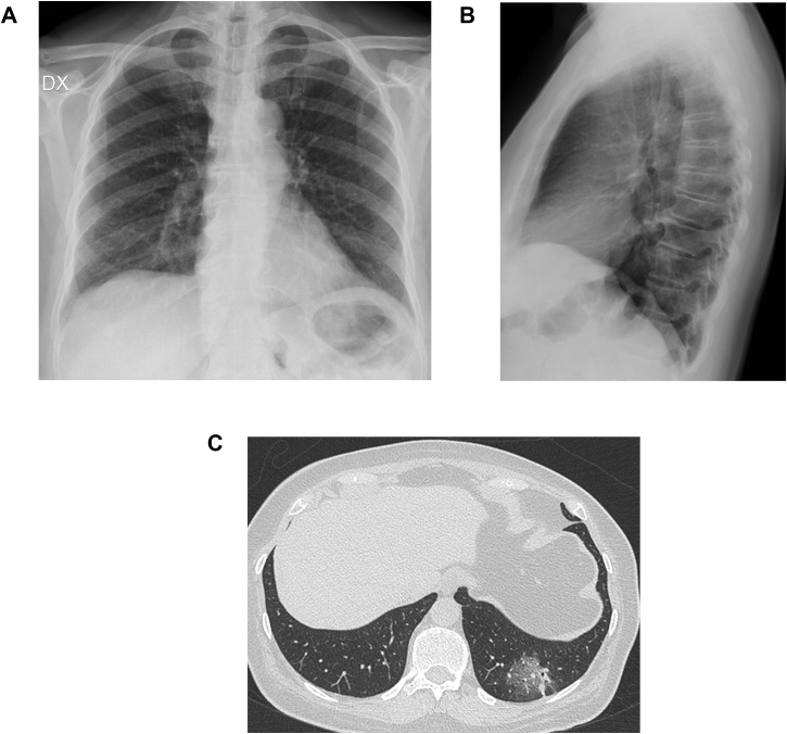
Due to the wide availability, rapid execution, low cost, and possibility of being acquired at the patient's bed, chest X-Ray is a fundamental tool in the diagnosis, follow-up and evaluation of the treatment effectiveness of patients with pneumonia, also in the context of COVID-19 infection. However, false negative cases are possible. We report 4 cases of false negative chest X-Rays, in patients who were diagnosed positive for COVID-19 by real-time transverse-transcript-polymerase chain reaction (RT-PCR), and executed chest unenhanced CTs just after the X-Rays, demonstrating signs of COVID-19 pneumonia.

Keywords: COVID-19; Multidetector computed tomography; Pneumonia; Radiography; Severe acute respiratory syndrome coronavirus 2; Viral.


Conflict of interest statement

Conflict of interest statement None.

Figures

Figure 1
Figure 1
Posteroanterior (Fig. 1A) and lateral (Fig. 1B) projections of the chest X-Ray of a thirty-seven-year-old male colleague, who presented to our Emergency Department with cough and fever up to 39° for 2 days. Chest X-Ray did not demonstrate any lung abnormalities. Chest CT executed just after the X-Ray, showed the presence of a crazy paving area in the right posterior costophrenic recess with mild pleural effusion (Fig. 1C). A small subpleural area of GGO is located anteriorly in the upper right lobe (Fig. 1D).
Figure 2
Figure 2
Posteroanterior (Fig. 2A) and lateral (Fig. 2B) projections of the chest X-Ray of a thirty-seven-year-old female colleague, who presented with cough and fever. Chest X-Ray did not demonstrate any lung abnormalities. Chest CT executed just after the X-Ray, showed an area of crazy paving in the left posterior costophrenic recess (Fig. 2B); patchy GGOs are also bilaterally recognizable (Fig. 2B and C).
Figure 3
Figure 3
Posteroanterior (Fig. 3A) and lateral (Fig. 3B) projections of the chest X-Ray of a thirty-three-year-old nurse, who presented with cough and fever. Chest X-Ray was reported as negative; at a review, a slight thickening of the bronchovascular bundles could be observed at the lower right field. Chest CT executed just after the X-Ray, showed an extensive consolidation, with a peripheral posterior location in the right lower lobe, and some patchy GGOs with lateral peripheral distribution (Fig. 3C).
Figure 4
Figure 4
Posteroanterior (Fig. 4A) and lateral (Fig. 4B) projections of the chest X-Ray of a fifty-six-year-old woman, who presented with chest pain, cough, dyspnoea, and fever. Chest CT demonstrated an area of crazy paving pattern, posteriorly located in the lower left lobe (Fig. 4C).

References

    1. Wong H.Y.F., Lam H.Y.S., Fong A.H., Leung S.T., Chin T.W., Lo C.S.Y. Frequency and distribution of chest radiographic findings in COVID-19 positive patients. Radiology. 2019;27:201160. doi: 10.1148/radiol.2020201160. - DOI - PMC - PubMed
    1. Zhou S., Wang Y., Zhu T., Xia L. CT features of coronavirus disease 2019 (COVID-19) pneumonia in 62 patients in Wuhan, China. Am J Roentgenol. 2020:1–8. doi: 10.2214/AJR.20.22975. - DOI - PubMed
    1. Ng M.-Y., Lee E.Y.P., Yang J., Li X., Wang H., Mei-Sze M. Imaging profile of the COVID-19 infection: radiologic findings and literature review. Radiol Cardiothorac Imag. 2020;2(1) doi: 10.1148/ryct.2020200034. - DOI - PMC - PubMed
    1. Chung M., Bernheim A., Mei X., Zhang N., Zeng X., Cui J. CT imaging features of 2019 novel coronavirus (2019-nCoV) Radiology. 2020 doi: 10.1148/radiol.2020200230. - DOI - PMC - PubMed
    1. Ai T., Yang Z., Hou H., Zhan C., Chen C., Lv W. Correlation of chest CT and RT-PCR testing in coronavirus disease 2019 (COVID-19) in China: a report of 1014 cases. Radiology. 2020 doi: 10.1148/radiol.2020200642. - DOI - PMC - PubMed

Publication types

MeSH terms