. 2020 Jul 10;295(28):9531-9541.
doi: 10.1074/jbc.RA120.013354. Epub 2020 May 18.

Molecular mechanism of the recognition of bacterially cleaved immunoglobulin by the immune regulatory receptor LILRA2

Affiliations

Molecular mechanism of the recognition of bacterially cleaved immunoglobulin by the immune regulatory receptor LILRA2

Rika Yamazaki et al. J Biol Chem. .

Abstract

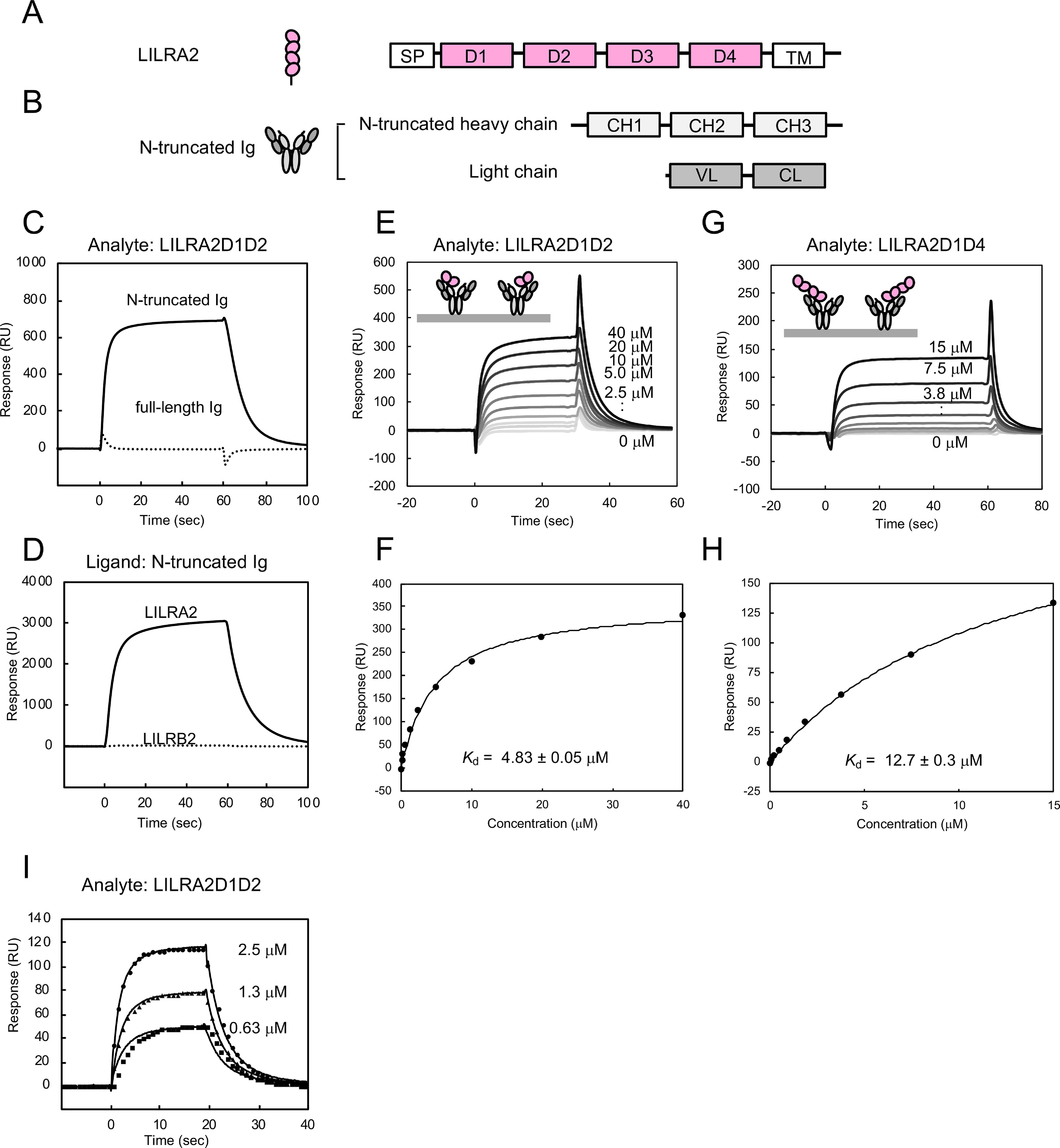
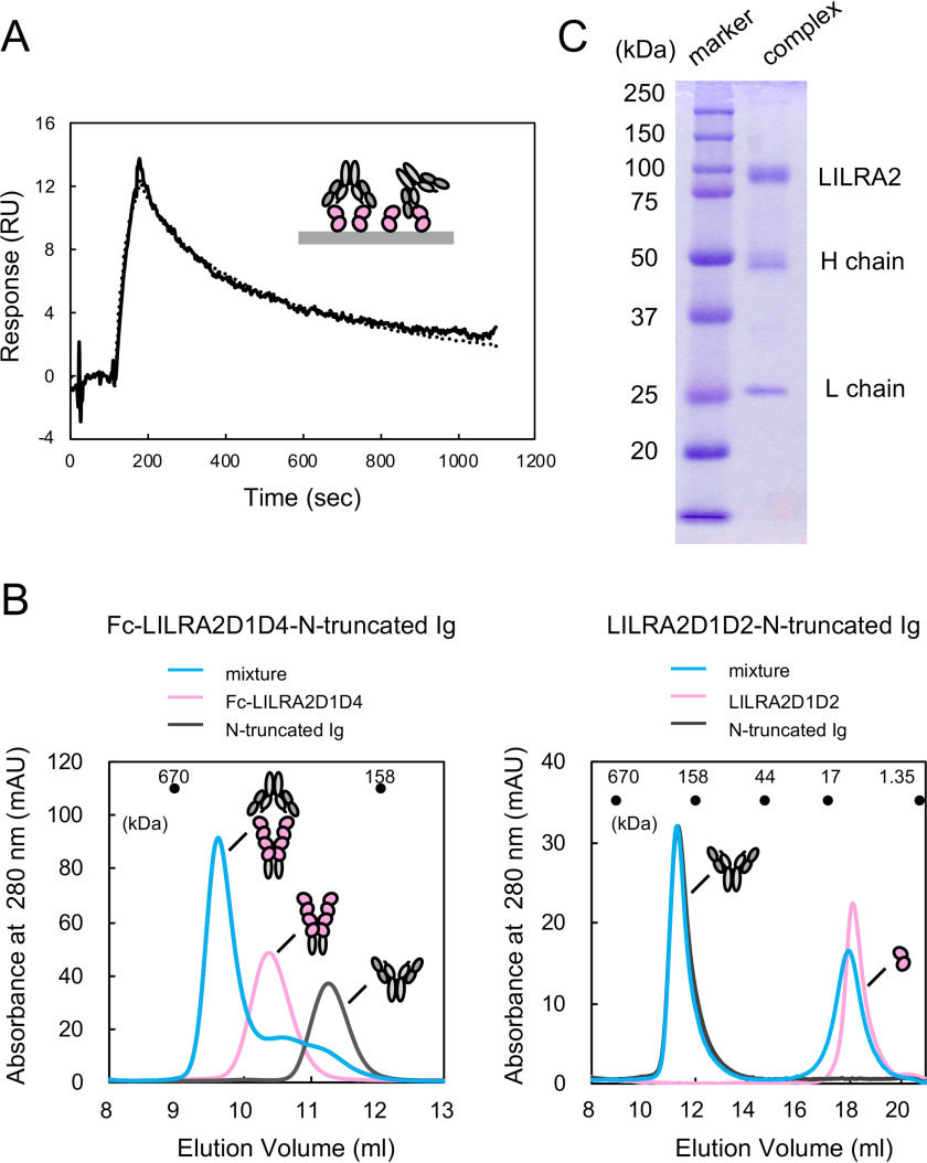
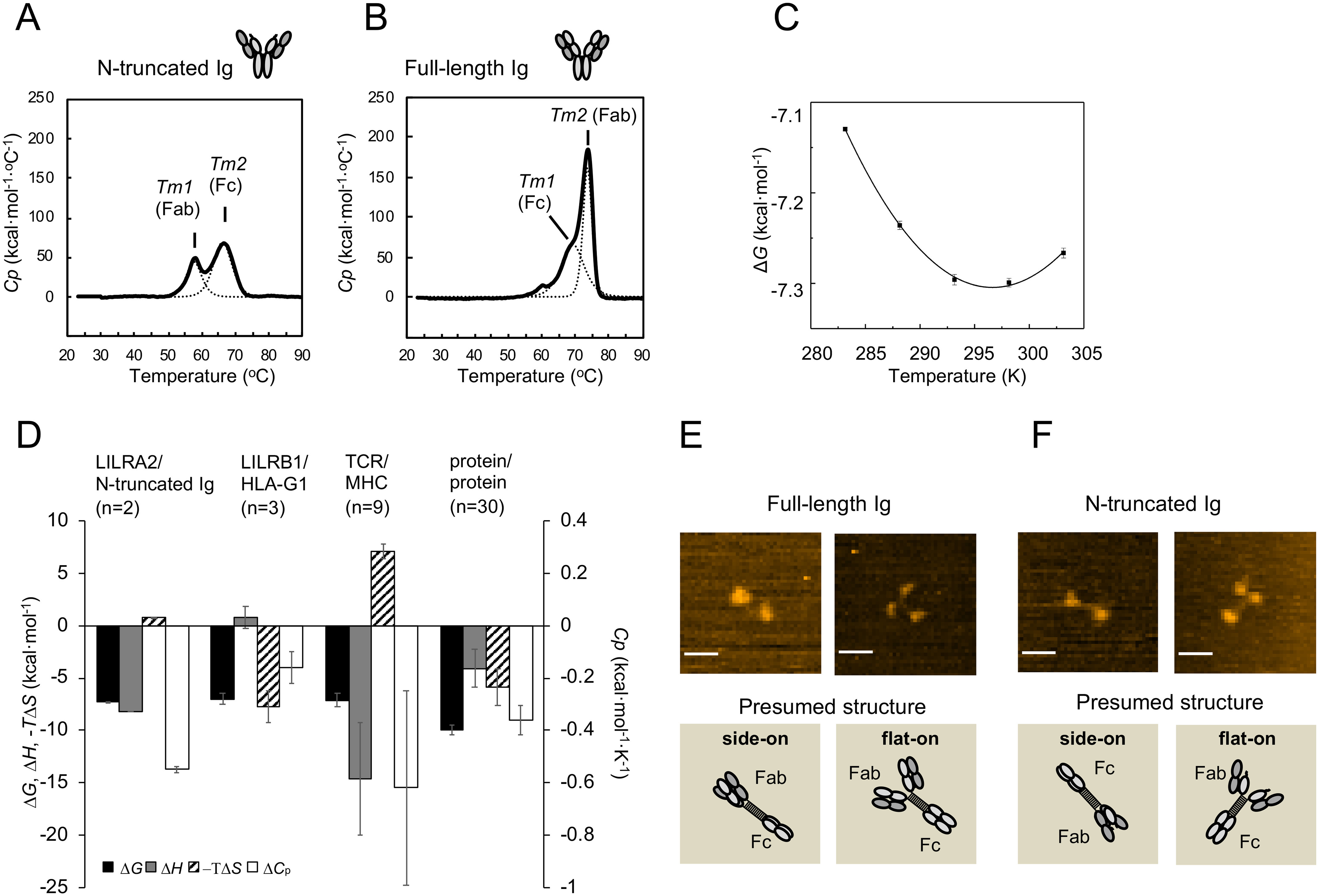
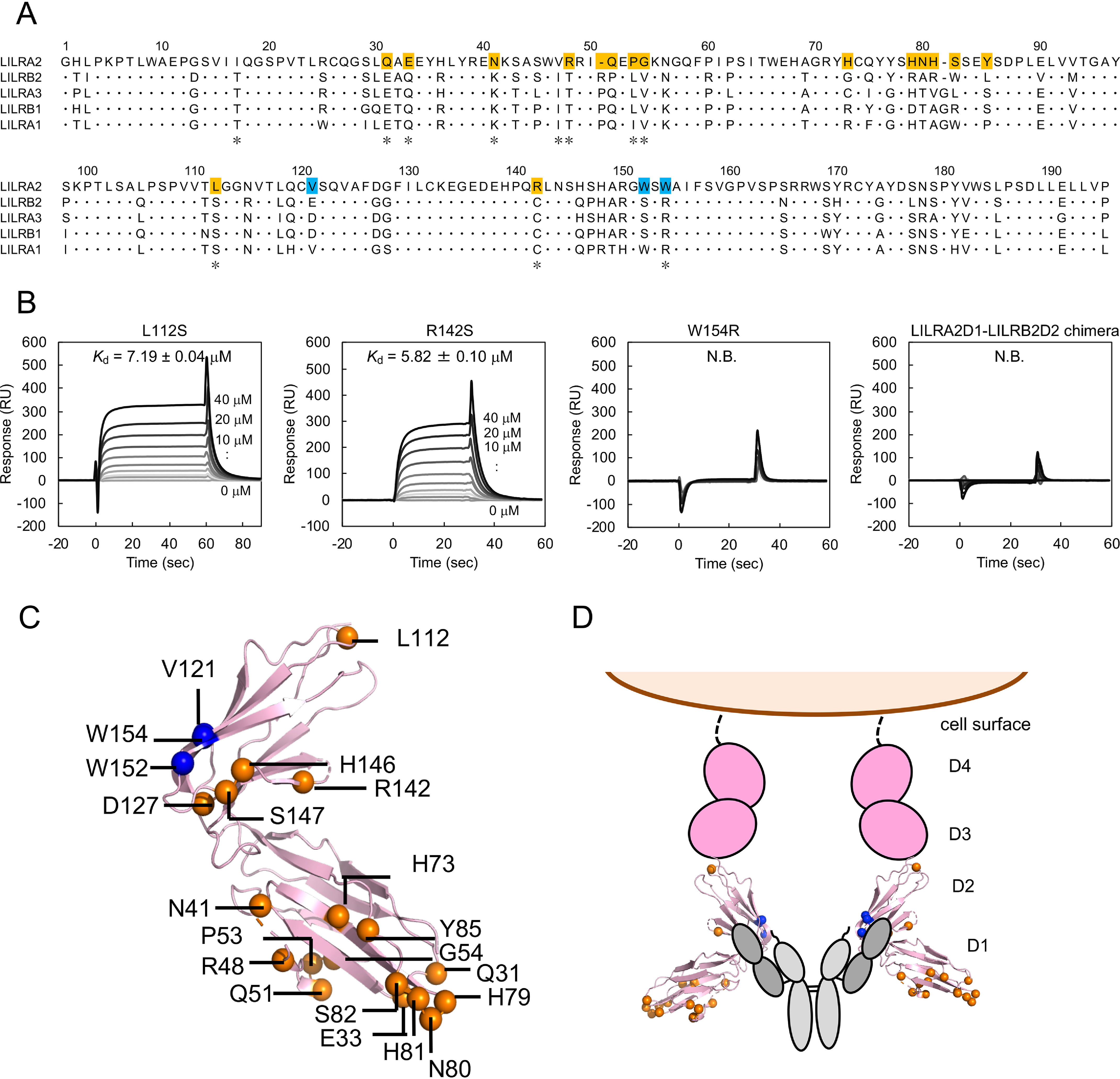
Human leukocyte immunoglobulin-like receptors (LILRs) typically regulate immune activation by binding to the human leukocyte antigen class I molecules. LILRA2, a member of the LILR family, was recently reported to bind to other unique ligands, the bacterially degraded Igs (N-truncated Igs), for the activation of immune cells. Therefore, LILRA2 is currently attracting significant attention as a novel innate immune receptor. However, the detailed recognition mechanisms required for this interaction remain unclear. In this study, using several biophysical techniques, we uncovered the molecular mechanism of N-truncated Ig recognition by LILRA2. Surface plasmon resonance analysis disclosed that LILRA2 specifically binds to N-truncated Ig with weak affinity (Kd = 4.8 μm) and fast kinetics. However, immobilized LILRA2 exhibited a significantly enhanced interaction with N-truncated Ig due to avidity effects. This suggests that cell surface-bound LILRA2 rapidly monitors and identifies bi- or multivalent abnormal N-truncated Igs through specific cross-linking to induce immune activation. Van't Hoff analysis revealed that this interaction is enthalpy-driven, with a small entropy loss, and results from differential scanning calorimetry indicated the instability of the putative LILRA2-binding site, the Fab region of the N-truncated Ig. Atomic force microscopy revealed that N truncation does not cause significant structural changes in Ig. Furthermore, mutagenesis analysis identified the hydrophobic region of LILRA2 domain 2 as the N-truncated Ig-binding site, representing a novel ligand-binding site for the LILR family. These results provide detailed insights into the molecular regulation of LILR-mediated immune responses targeting ligands that have been modified by bacteria.

Keywords: LILRA2; atomic force microscopy (AFM); bacterially cleaved immunoglobulin; cell surface receptor; immune regulation; immune regulatory receptor; immunoglobulin G (IgG); immunology; leukocyte immunoglobulin-like receptor (LILR); protein-protein interaction; surface plasmon resonance (SPR).


Conflict of interest statement

Conflict of interest—The authors declare that they have no conflicts of interest with the contents of this article.

Figures

Figure 1.
Figure 1.
Binding analysis between LILRA2 and N-truncated Ig. A, Schematic structures of LILRA2. SP, signal peptide; D, domain; TM, transmembrane domain. B, Schematic structures of N-truncated Ig. C, The SPR sensorgrams of injection of LILRA2D1D2 over N-truncated Ig (solid lines) or full-length Ig (dotted lines) are shown. D, Interaction analysis of LILRA2D1D2 or LILRB2D1D2 with N-truncated Ig. N-truncated Ig was immobilized on a sensor chip at 20,000 RU. 9 μm LILRA2D1D2 or LILRB2D1D2 was injected as the analyte. E, SPR sensorgram of continuous injection of LILRA2D1D2 at various concentrations, from 0.16 μm to 40 μm, over N-truncated Ig immobilized sensor chip. F, Equilibrium analysis of panel E. The black line represents nonlinear fits of the 1:1 Langmuir binding fitting. Kd value is presented as mean ± standard deviation from three experiments. G, SPR sensorgram of continuous injection of LILRA2D1D4 at various concentrations, from 0.12 μm to 15 μm, over N-truncated Ig immobilized sensor chip. H, Equilibrium analysis of panel G. The black line represents nonlinear fits of the 1:1 Langmuir binding fitting. Kd value is presented as mean ± range from two experiments. I, The kinetic analysis of LILRA2 binding to N-truncated Ig. SPR sensorgram of LILRA2 at various concentrations, from 0.63 μm to 2.5 μm, over N-truncated Ig immobilized sensor chip, is shown. Circle, triangle, and square show the fitting curve based on the global fitting analysis using the 1:1 Langmuir binding model.
Figure 2.
Figure 2.
Bivalent effect of the interaction between LILRA2 and N-truncated Ig. A, 47 nm N-truncated Ig was injected over the LILRA2D1D2 immobilized (100 RU) sensor chip. The black solid line represents raw data. The dotted line represents the fitting curve based on the bivalent analyte model. The ka1, kd1, ka2, and kd2 of the interaction were determined by bivalent fitting to be 1.6 (105 M−1·s−1), 0.080 (s−1), 0.35 (RU−1·s−1), and 0.094 (s−1), respectively. B, The size exclusion chromatogram of the mixture of Fc fusion LILRA2D1D4 or LILAR2D1D2 and N-truncated Ig (blue line). The chromatogram of free Fc fusion LILRA2D1D4 or LILRA2D1D2 and N-truncated Ig are also shown as a pink line and gray line, respectively. Approximate positions of molecular weight standards are shown above the chromatogram. C, SDS-PAGE of the eluted fraction of the complex of Fc fusion LILRA2D1D4 and N-truncated Ig from size exclusion chromatography of panel B.
Figure 3.
Figure 3.
Thermodynamic features of LILRA2/N-truncated Ig interaction and structural characteristics of N-truncated Ig. A–B, DSC analyses of N-truncated Ig (A) and full-length Ig (B) are shown. Solid lines represent raw data. Dotted lines represent fitting curves. C, Thermodynamic analysis of LILRA2 binding to N-truncated Ig. The interaction was measured at several temperatures (10–30 °C) and converted into the standard free energy of binding (ΔG). Values for the enthalpic (ΔH) and standard entropic (–TΔS) changes (at 25 °C) and the specific heat capacity (ΔCp) were calculated by fitting the nonlinear form of the van't Hoff equation to these data (see Experimental procedures). Error bars show the range from two independent experiments. D, Comparison of thermodynamic properties (at 25 °C) of several cell surface intermolecular interactions. Data for LILRB1/HLA-G1 and TCR/peptide–MHC interactions are from references and , , respectively. Data for protein–protein interactions (excluding antibody–antigen interactions) are from reference . The mean and standard error of 30 types of protein–protein interactions are depicted. E–F, Representative HS-AFM images of full-length Ig (E) and N-truncated Ig (F) (scale bars, 25 nm). The images show full-length Ig and N-truncated Ig absorbed on mica in different orientations. Schematic images corresponding to the above HS-AFM images show presumed absorbed protein structure. The upper right image of panel F was selected from Fig. S3 as a representative of the flat-on structure.
Figure 4.
Figure 4.
Mutagenesis analysis for LILRA2 binding mode toward N-truncated Ig. A, Alignment of group 1 LILRs. The asterisks below the sequence indicate the LILRA2 unique residues among group 1 LILRs. The mutated residues that did not affect N-truncated Ig binding are shown with orange background. The identified residues involved in ligand binding are shown with blue background. B, Mutagenesis analysis using SPR. LILRA2 mutants were injected onto N-truncated Ig immobilized chip. Kd value is presented as mean ± range from two experiments or mean ± standard deviation from three experiments. N.B. means no detectable binding. C, Mapping of mutated amino acid residues of LILRA2. Blue spheres represent the residues that are involved in N-truncated Ig binding. Orange spheres represent the residues that are not involved in N-truncated Ig binding. The structure was based on LILRB2 (PDB entry 2GW5). D, Proposed model of N-truncated Ig recognition by LILRA2.

Similar articles

Cited by

References

    1. Kuroki K., Furukawa A., and Maenaka K. (2012) Molecular recognition of paired receptors in the immune system. Front. Microbiol. 3, 429 10.3389/fmicb.2012.00429 - DOI - PMC - PubMed
    1. Sharma P., and Allison J. P. (2015) Immune checkpoint targeting in cancer therapy: toward combination strategies with curative potential. Cell 161, 205–214 10.1016/j.cell.2015.03.030 - DOI - PMC - PubMed
    1. Kang X., Kim J., Deng M., John S., Chen H., Wu G., Phan H., and Zhang C. C. (2016) Inhibitory leukocyte immunoglobulin-like receptors: immune checkpoint proteins and tumor sustaining factors. Cell Cycle 15, 25–40 10.1080/15384101.2015.1121324 - DOI - PMC - PubMed
    1. Shiroishi M., Tsumoto K., Amano K., Shirakihara Y., Colonna M., Braud V. M., Allan D. S., Makadzange A., Rowland-Jones S., Willcox B., Jones E. Y., van der Merwe P. A., Kumagai I., and Maenaka K. (2003) Human inhibitory receptors Ig-like transcript 2 (ILT2) and ILT4 compete with CD8 for MHC class I binding and bind preferentially to HLA-G. Proc. Natl. Acad. Sci. U S A 100, 8856–8861 10.1073/pnas.1431057100 - DOI - PMC - PubMed
    1. Chapman T. L., Heikeman A. P., and Bjorkman P. J. (1999) The inhibitory receptor LIR-1 uses a common binding interaction to recognize class I MHC molecules and the viral homolog UL18. Immunity 11, 603–613 10.1016/S1074-7613(00)80135-1 - DOI - PubMed

Publication types

MeSH terms

Substances

Associated data

LinkOut - more resources