. 2020 Oct;27(10):2952-2972.
doi: 10.1038/s41418-020-0554-8. Epub 2020 May 18.

The proteasome activator REGγ accelerates cardiac hypertrophy by declining PP2Acα-SOD2 pathway

Affiliations

The proteasome activator REGγ accelerates cardiac hypertrophy by declining PP2Acα-SOD2 pathway

Yifan Xie et al. Cell Death Differ. 2020 Oct.

Abstract

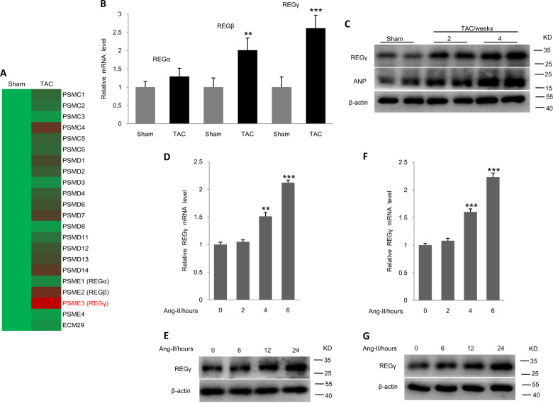
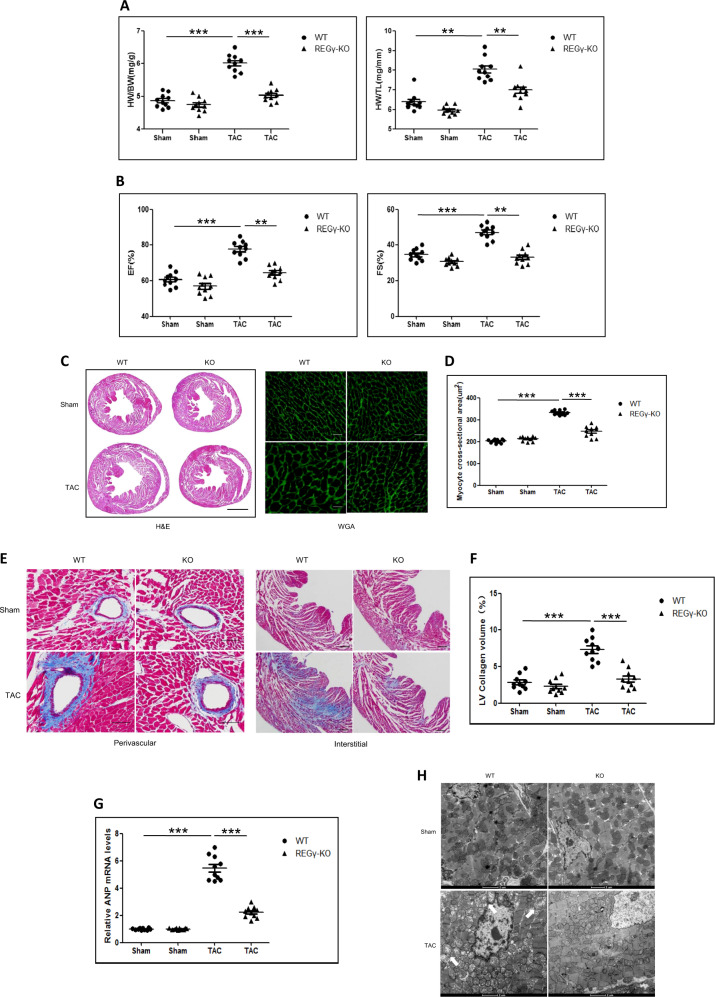
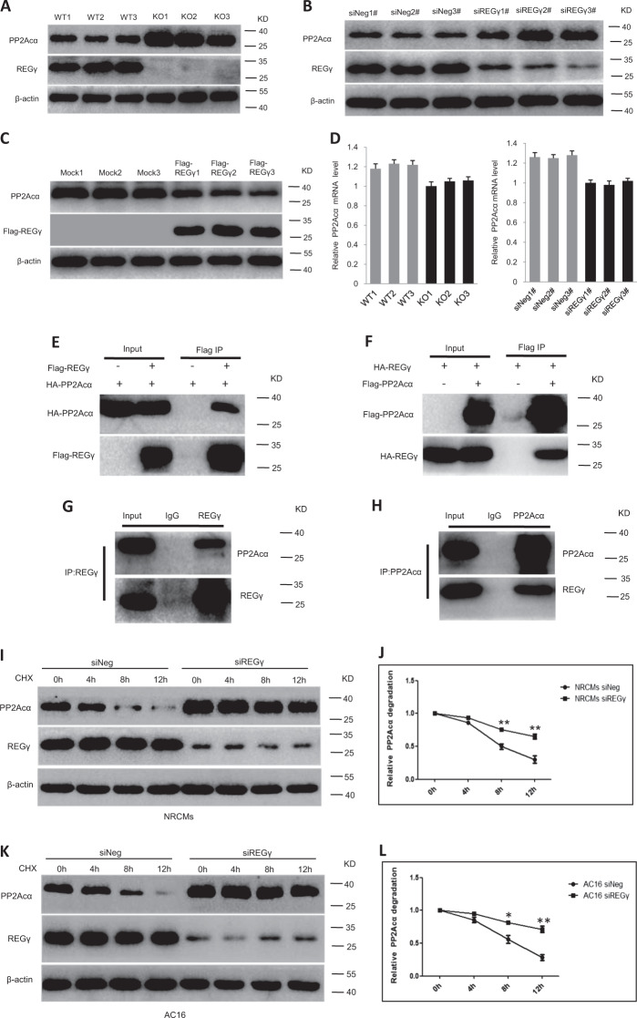
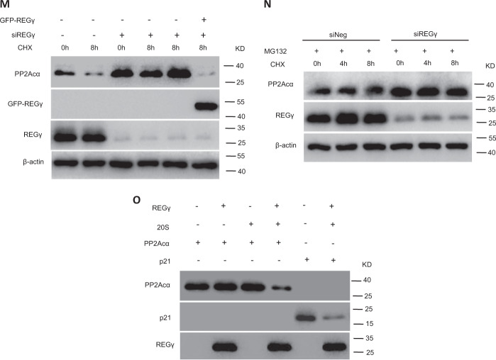
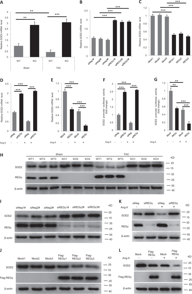
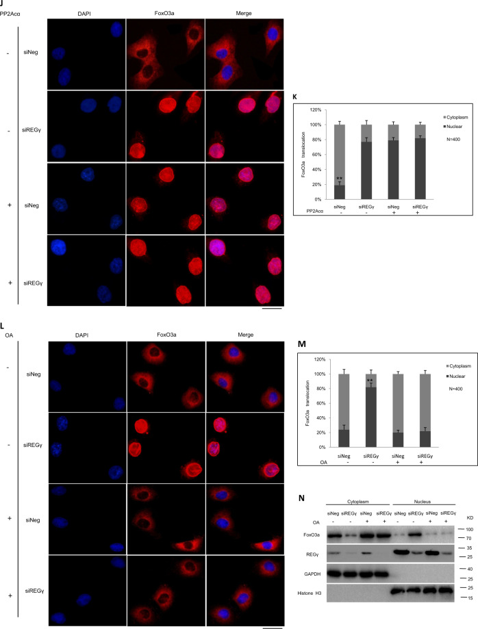
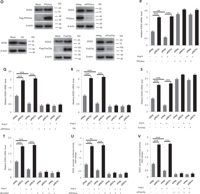
Pathological cardiac hypertrophy eventually leads to heart failure without adequate treatment. REGγ is emerging as 11S proteasome activator of 20S proteasome to promote the degradation of cellular proteins in a ubiquitin- and ATP-independent manner. Here, we found that REGγ was significantly upregulated in the transverse aortic constriction (TAC)-induced hypertrophic hearts and angiotensin II (Ang II)-treated cardiomyocytes. REGγ deficiency ameliorated pressure overload-induced cardiac hypertrophy were associated with inhibition of cardiac reactive oxygen species (ROS) accumulation and suppression of protein phosphatase 2A catalytic subunit α (PP2Acα) decay. Mechanistically, REGγ interacted with and targeted PP2Acα for degradation directly, thereby leading to increase of phosphorylation levels and nuclear export of Forkhead box protein O (FoxO) 3a and subsequent of SOD2 decline, ROS accumulation, and cardiac hypertrophy. Introducing exogenous PP2Acα or SOD2 to human cardiomyocytes significantly rescued the REGγ-mediated ROS accumulation of Ang II stimulation in vitro. Furthermore, treatment with superoxide dismutase mimetic, MnTBAP prevented cardiac ROS production and hypertrophy features that REGγ caused in vivo, thereby establishing a REGγ-PP2Acα-FoxO3a-SOD2 pathway in cardiac oxidative stress and hypertrophy, indicates modulating the REGγ-proteasome activity may be a potential therapeutic approach in cardiac hypertrophy-associated disorders.


Conflict of interest statement

The authors declare that they have no conflict of interest.

Figures

Fig. 1
Fig. 1. REGγ is increased in hypertrophic hearts.
a Profile of the proteasome activators (subunits) expression in Sham or TAC-operated mouse heart at 4 weeks. b Relative fold change of REGα, REGβ, and REGγ mRNA expression in Sham or TAC-operated mouse heart at 4 weeks by RT-qPCR analysis (n = 6 per group, **P < 0.01, ***P < 0.001, Student’s t test). Protein levels of REGγ in mouse heart of Sham or TAC operation after 2 or 4 weeks (c) by immunoblotting analysis. mRNA and protein levels of REGγ in (d, e) NRCMs and (f, g) AC16 cells exposed to Ang II at different time points by RT-qPCR and immunoblotting analysis. (The experiments were repeated three times; error bars represent standard deviation, **P < 0.01, ***P < 0.001, Student’s t test).
Fig. 2
Fig. 2. REGγ deficiency ameliorates TAC-induced cardiac hypertrophy phenotypes.
a Heart weight to body weight (HW/BW) ratio and heart weight to tibia length (HW/TL) ratio. b Heart ejection fraction (EF) and fractional shortening (FS). c Heart haematoxylin and eosin (H&E) staining (scale bars: 2 mm). Wheat germ agglutinin staining (scale bars: 40 μm) and d corresponding quantitation graphs of myocyte cross-sectional area. e Masson staining to detect fibrosis of heart collagen (scale bars: 100 μm) and f corresponding quantitation graphs of heart fibrosis, and g heart ANP mRNA expression, and h representative transmission electron microscopy (TEM) images of the WT and REGγ-KO mice after sham or TAC operation for 4 weeks (n = 10 for each genotype; **P < 0.01, ***P < 0.001, one-way ANOVA test).
Fig. 3
Fig. 3. REGγ deficiency improves cardiac oxidative stress in response to hypertrophic stimuli.
a REGγ deficiency inhibits cardiac ROS accumulation (scale bars: 50 μm) and b corresponding quantitation graphs of DHE relative fluorescence in mice after TAC operation for 4 weeks by DHE staining analysis (n = 10 for each genotype; *** P < 0.001, one-way ANOVA test). REGγ knockdown inhibits ROS accumulation (scale bars: 40 μm) and corresponding quantitation bar graphs of DHE relative fluorescence in (c, d) NRCMs and (e, f) human cardiomyocyte AC16 cells after Ang II treatment by DHE staining analysis. (The experiments were repeated three times; error bars represent standard deviation, **P < 0.01, ***P < 0.001, one-way ANOVA test).
Fig. 4
Fig. 4. REGγ interacts with PP2Acα and directs its degradation.
a REGγ knockout increases of PP2Acα protein levels in mice heart, and b REGγ knockdown upregulates PP2Acα protein levels, and c REGγ overexpression downregulates PP2Acα protein levels in AC16 cells by immunoblotting analysis, whereas d REGγ knockout or knockdown downregulates PP2Acα mRNA expression in murine heart or AC16 cells. e Interaction between REGγ and PP2Acα in 293T cells was determined by coimmunoprecipitation and immunoblotting analysis following transient transfection of Flag-REGγ/Flag vector and HA-PP2Acα into 293T cells. f Reciprocal interaction between REGγ and PP2Acα was performed by coimmunoprecipitation as indicated following transient transfection of Flag-PP2Acα/Flag vector and HA-REGγ into 293T cells. g Endogenous REGγ in AC16 cells was precipitated using anti-REGγ antibody or with IgG control, and coprecipitated PP2Acα was detected by immunoblotting. h Reciprocal interaction between endogenous REGγ and PP2Acα in AC16 cells was performed by using anti-PP2Acα antibody or IgG control, and coprecipitated REGγ was detected by immunoblotting. Stability of endogenous PP2Acα in (i) siNeg and siREGγ NRCMs and (j) corresponding quantitation graphs of relative PP2Acα degradation, or (k) siNeg and siREGγ AC16 cells and (l) corresponding quantitation graphs of relative PP2Acα degradation. (The experiments were repeated three times; error bars represent standard deviation, *P < 0.05, **P < 0.01, Student’s t test.) Cells were treated with CHX (100 μg/mL) for indicated times followed by immunoblotting. Stability of endogenous PP2Acα in (m) siNeg, siREGγ, and siREGγ plus GFP-REGγ plasmid AC16 cells and (n) AC16 cells pretreated with MG132. Cells were treated with CHX (100 μg/mL) for indicated times followed by immunoblotting. o REGγ directly promoted the degradation of PP2Acα. In vitro proteolytic analyses were performed using purified REGγ, 20S proteasome, and in vitro translated PP2Acα protein as indicated and described in “Materials and methods.” A known substrate of REGγ, p21, was shown as a positive control.
Fig. 5
Fig. 5. REGγ declines SOD2 expression.
a REGγ deficiency inhibits the decline in cardiac SOD2 mRNA expression of mice in response to TAC operation for 4 weeks (n = 6 per group; **P < 0.01, ***P < 0.001, one-way ANOVA test). b REGγ knockdown upregulates SOD2 mRNA expression, and c REGγ overexpression downregulates SOD2 mRNA expression in human cardiomyocyte AC16 cells. (The experiments were repeated three times; error bars represent standard deviation, **P < 0.01, ***P < 0.001, one-way ANOVA test.) d REGγ knockdown inhibits the decline in SOD2 mRNA expression, and e REGγ overexpression promotes the decline in SOD2 mRNA expression in human cardiomyocyte AC16 cells in response to Ang II treatment. f, g Similar results were observed in AC16 cells by SOD2 promoter luciferase assays. (The experiments were repeated three times; error bars represent standard deviation, **P < 0.01, ***P < 0.001, one-way ANOVA test.) Similar consistent results were also observed by immunoblotting (h) in mice heart of sham or TAC operation for 4 weeks, and immunoblotting (il) in AC16 cells at indicated treatment.
Fig. 6
Fig. 6. REGγ declines SOD2 expression in a PP2Acα–FoxO3a-dependent manner.
a Protein levels of PP2Acα, P-FoxO3a, and SOD2 in REGγ+/+ and REGγ−/− heart tissue, and siNeg and siREGγ NRCMs and AC16 cells. Knocking out or silencing REGγ decreases FoxO3a phosphorylation levels in (b) murine heart tissue or in (c) human cardiomyocyte AC16 cells. REGγ knockdown promoted the translocation of FoxO3a from the cytoplasm to the nucleus in AC16 cells by d immunofluorescence analysis (scale bars: 20 μm) and e corresponding quantitation graphs, and f cell fractionation assay. (The experiments were repeated three times. Error bars represent standard deviation, **P < 0.01, Student’s t test.) g The levels of P-FoxO3a after OA treatment or PP2Acα overexpression in AC16 cells. h Overexpressing PP2Acα or i blocking PP2Acα activity by OA treatment significantly diminished the change of FoxO3a phosphorylation levels which were caused by REGγ knockdown in AC16 cells, similar results of (jm) immunofluorescence analysis (scale bars: 20 μm) and corresponding quantitation graphs of FoxO3a translocation, and n cell fractionation assay was observed. (The experiments were repeated three times. Error bars represent standard deviation, **P < 0.01, one-way ANOVA test.) o Expression of SOD2 after PP2Acα overexpression, knockdown or activity inhibition by PP2Acα plasmid, siPP2Acα transfection or OA treatment, or FoxO3a overexpression or knockdown by FoxO3a or siFoxO3a plasmid transfection. pr PP2Acα knockdown, overexpression, or activity inhibition dramatically rescued or attenuated SOD2 mRNA expression of Ang II stimuli regardless of REGγ levels. s, t FoxO3a overexpression or knockdown diminished the change of SOD2 mRNA expression of Ang II stimuli which REGγ caused. u, v Similar results of luciferase assays were observed in AC16 cells. (The experiments were repeated three times; error bars represent standard deviation, **P < 0.01, ***P < 0.001, one-way ANOVA test).
Fig. 7
Fig. 7. REGγ-induced cardiac ROS production and hypertrophy-related anomalies depend on PP2Acα–SOD2 axis.
PP2Acα indeed requires SOD2 for the inhibition ROS accumulation in cardiomyocyte. a, b SOD2 overexpression inhibited ROS accumulation caused by PP2Acα knockdown, and c, d PP2Acα or e, f SOD2 overexpression rescued ROS accumulation caused by REGγ in Ang II-treated human cardiomyocyte AC16 cells by DHE staining (scale bars: 40 μm) and corresponding quantitation graphs of DHE relative fluorescence, indicating PP2Acα–SOD2 axis essentially contributes to the effects of REGγ on cardiac ROS accumulation. (The experiments were repeated three times; error bars represent standard deviation, ***P < 0.01, one-way ANOVA test.) MnTBAP (MnT) treatment prevented cardiac ROS production and hypertrophy-related anomalies that caused by REGγ. g Heart DHE staining (scale bars: 50 μm) and h corresponding quantitation graphs of DHE relative fluorescence, i heart weight to body weight (HW/BW) ratio and heart weight to tibia length (HW/TL) ratio, j heart ejection fraction (EF) and fractional shortening (FS), k heart haematoxylin and eosin (H&E) staining (scale bars: 40 μm), and (l) corresponding quantitation graphs of myocyte cross-sectional area, m heart ANP mRNA expression of the WT and REGγ-KO mice after indicated operation (Sham-MnT, TAC, or TAC-MnT) for 4 weeks (n = 6 for each genotype; ***P < 0.001, **P < 0.01, one-way ANOVA test).

References

    1. Frey N, Olson EN. Cardiac hypertrophy: the good, the bad, and the ugly. Annu Rev Physiol. 2003;65:45–79. - PubMed
    1. Korolchuk VI, Menzies FM, Rubinsztein DC. Mechanisms of cross-talk between the ubiquitin-proteasome and autophagy-lysosome systems. FEBS Lett. 2010;584:1393–8. - PubMed
    1. Mizushima N, Komatsu M. Autophagy: renovation of cells and tissues. Cell. 2011;147:728–41. - PubMed
    1. Cacciapuoti F. Role of ubiquitin-proteasome system (UPS) in left ventricular hypertrophy (LVH) Am J Cardiovasc Dis. 2014;4:1–5. - PMC - PubMed
    1. Varshavsky A. Regulated protein degradation. Trends Biochem Sci. 2005;30::283–6. - PubMed

Publication types