Review
. 2020 May;36(3):183-194.
doi: 10.6515/ACS.202005_36(3).20200213A.

Endothelial-to-Mesenchymal Transition in Calcific Aortic Valve Disease

Affiliations
Review

Endothelial-to-Mesenchymal Transition in Calcific Aortic Valve Disease

Xiaochun Ma et al. Acta Cardiol Sin. 2020 May.

Abstract

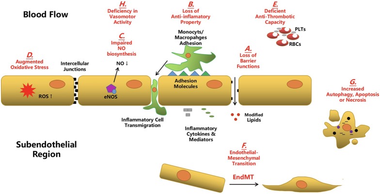
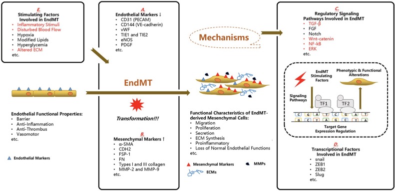
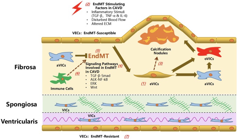
Calcific aortic valve disease (CAVD) represents a significant threat to cardiovascular health worldwide, and the incidence of this sclerocalcific valve disease has rapidly increased along with a rise in life expectancy. Compelling evidence has suggested that CAVD is an actively and finely regulated pathophysiological process even though it has been referred to as "degenerative" for decades. A striking similarity has been noted in the etiopathogenesis between CAVD and atherosclerosis, a classical proliferative sclerotic vascular disease.1 Nevertheless, pharmaceutical trials that attempted to target inflammation and dyslipidemia have produced disappointing results in CAVD. While senescence is a well-documented risk factor, the sophisticated regulatory networks have not been adequately explored underlying the aberrant calcification and osteogenesis in CAVD. Valvular endothelial cells (VECs), a type of resident effector cells in aortic leaflets, are crucial in maintaining valvular integrity and homeostasis, and dysfunctional VECs are a major contributor to disease initiation and progression. Accumulating evidence suggests that VECs undergo a phenotypic and functional transition to mesenchymal or fibroblast-like cells in CAVD, a process known as the endothelial-to-mesenchymal transition (EndMT) process. The relevance of this transition in CAVD has recently drawn great interest due to its importance in both valve genesis at an embryonic stage and CAVD development at an adult stage. Hence EndMT might be a valuable diagnostic and therapeutic target for disease prevention and treatment. This mini-review summarized the relevant literature that delineates the EndMT process and the underlying regulatory networks involved in CAVD.

Keywords: Calcific aortic valve disease; Endothelial-to-mesenchymal transition; Regulatory network; Valvular endothelial cell.


Figures

Figure 1
Figure 1
Aberrant endothelial cells in atherosclerosis and calcific aortic valve disease. During the development of atherosclerosis and calcific aortic valve disease (CAVD), aberrant endothelial cells (ECs) disturb markedly the micro-environmental homeostasis of cardiovascular system and contribute greatly to the disease progression. (A) Endothelial dysfunction leads to the loss of barrier function and results in the infiltration of modified lipids to the subendothelial region. (B) Dysfunctional ECs transfer from an anti-inflammatory phenotype to a pro-inflammatory phenotype. Particularly, an increased expression of adhesion molecules and pro-inflammatory cytokines and mediators by these ECs promote the adhesion and subsequent transmigration of circulating immune cells to the subendothelial region. (C) The bio-synthesis and release of nitric oxide (NO) is compromised in the dysfunctional ECs and it impedes the physiological functions of ECs. (D) An array of pathological environmental stimuli induces the augmented oxidative stress in the aberrant ECs. (E) The anti-thrombotic property of ECs is deficient in the dysfunctional ECs. (F) Accumulating evidence suggests that the aberrant ECs undergo the endothelial-to-mesenchymal transition (EndMT) during which these ECs co-express the endothelial and mesenchymal markers and acquire the mesenchymal phenotypic and functional properties. (G) Persistent or potent pathological stimulators cause the increased level of autophagy and apoptosis and even the abnormal necrosis of ECs. (H) Dysfunctional ECs malfunction to regulate properly the vasomotor activity.
Figure 2
Figure 2
Endothelial-to-mesenchymal transition in the cardiovascular diseases. It has gained increasing attention that endothelial cells (ECs) experience the endothelial-to-mesenchymal transition (EndMT) in the evolution of common cardiovascular diseases including calcific aortic valve disease (CAVD) and this cellular reprogramming is potentially utilized as a novel diagnostic signature and therapeutic target. Under physiological conditions, ECs undertake a fundamental role in maintaining the homeostasis and integrity of cardiovascular architecture and their common duties include barrier, anti-inflammation, anti-thrombus and vasomotor. However, once the EndMT process is initiated, the normal functions of ECs are aberrantly interfered and progressively deprived. Furthermore, these transformed ECs gain the mesenchymal phenotypic characteristics such as proliferation, migration, secretion, extracellular matrix (ECM) synthesis and above all, pro-inflammatory capacities. In the process of EndMT, the cells co-expressing endothelial markers and mesenchymal markers is a hallmark. And in these cells, the listed endothelial markers (A) are down-regulated whereas the expression of mesenchymal markers (B) are enhanced. This cellular transition is launched by certain environmental stimuli, after which several well-documented signaling pathways (C) have been linked with the regulation of expression of certain target genes, on the transcriptional level by transcription factors (TF) (D), in most cases. (The red ones have been associated with the EndMT process in CAVD.) Several stimulators (E) have been correlated with the induction of EndMT. (The red ones have been reported in the EndMT process in CAVD). CD31, cluster of differentiation 31; CD144, cluster of differentiation 144; CDH2, N-cadherin; ECM, extracellular matrix; ECMs, extracellular matrixs; EndMT, endothelial-to-mesenchymal transition; eNOS, endothelial nitric oxide synthase; ERK, extracellular signal-regulated protein kinases; FGF, fibroblast growth factor; FN, fibronectin; FSP-1, fibroblast-specific protein-1; MMPs, matrix metalloproteinases; MMP-2, matrix metalloproteinase 2; MMP-9, matrix metalloproteinase 9; NF-kB, nuclear factor kappa-light-chain-enhancer of activated B cells; PDGF, platelet derived growth factor; PECAM, platelet endothelial cell adhesion molecule; TF1, transcription factor 1; TF2, transcription factor 2; TIE1, tyrosine kinase with immunoglobulin-like and EGF-like domains 1; TIE2, tyrosine kinase with immunoglobulin-like and EGF-like domains 2; VE-cadherin, vascular endothelial cadherin; vWF, von willebrand factor; ZEB1, zinc finger E-box-binding homeobox 1; ZEB2, zinc finger E-box-binding homeobox 2; α-SMA, alpha-smooth muscle actin; TGF-β, transforming growth factor-β.
Figure 3
Figure 3
Endothelial-to-mesenchymal transition in calcific aortic valve disease: the known knowledge and unresolved questions. The investigation on the role of endothelial-to-mesenchymal transition (EndMT) in calcific aortic valve disease has recently emerged. The evidence of EndMT in vivo has been found in the fibrosa layer (aortic aspect) of aortic valve, which was distal to the calcification nodule. In vitro evidence supported that inflammatory cytokines (including TNF-α, IL-6 and TGF-β), disturbed blood flow and altered extracellular matrix (ECM) was responsible for the induction of EndMT process, during which the TGF-β-Smad, ALK-NF-kB, ERK and Wnt signaling pathways might play a regulatory role. The transformed VECs (eVICs) might serve as an important reservoir for activated VICs (aVICs) whereas the EndMT process might be negatively regulated by VICs. eVICs might further transformed into an osteogenic type (oVIC) and cooperate with the VIC-derived oVICs to promote the CAVD progression. Several key unresolved questions are crucial in understanding the role of EndMT process in CAVS. (1) The origin of cells co-expressing endothelial and mesenchymal markers in the diseased aortic valve has to be further validated. (2) What are the effects of other risk factors/stimulating factors of CAVD on the EndMT process? (3) The relationship between EndMT process and calcific and osteogenic process remains to be explored in the future. In other words, it is elusive that how the EndMT process contributes to the different phases of CAVD from valve thickening, stenosis to calcification. (4) Several other well-known signaling pathways involved in the regulation of EndMT/EMT might also exert effects on the development of CAVD via EndMT. (5) Further in-depth investigation is required to verify a clearer correlation between the extent of EndMT, and severity and progression of CAVD based on pathological examination and echocardiography evaluation. (6) It is not clear how the interactions between VECs and other immune cells influence the EndMT process, and vice versa. (7) Last but not least, it is worthwhile of intensive efforts to verify the sub-population of EndMT-resistant VECs and the mechanisms modulating this obvious heterogeneity.

References

    1. Huang PH, Chen JW, Lin SJ. Effects of cardiovascular risk factors on endothelial progenitor cell. Acta Cardiol Sin. 2014;30:375–381. - PMC - PubMed
    1. Nkomo VT, Gardin JM, Skelton TN, et al. Burden of valvular heart diseases: a population-based study. Lancet. 2006;368:1005–1011. - PubMed
    1. Aikawa E, Libby P. A rock and a hard place: chiseling away at the multiple mechanisms of aortic stenosis. Circulation. 2017;135:1951–1955. - PMC - PubMed
    1. Duman H, Bahçeci I, Hamur H, et al. The relationship between serum apelin levels and the severity of calcific aortic stenosis. Acta Cardiol Sin. 2018;34:259–266. - PMC - PubMed
    1. Shen M, Tastet L, Bergler-Klein J, et al. Blood, tissue and imaging biomarkers in calcific aortic valve stenosis: past, present and future. Curr Opin Cardiol. 2018;33:125–133. - PubMed

LinkOut - more resources