. 2020 Apr 3:64.
doi: 10.29219/fnr.v64.3744. eCollection 2020.

A novel exopolysaccharide produced by Lactobacillus coryniformis NA-3 exhibits antioxidant and biofilm-inhibiting properties in vitro

Affiliations

A novel exopolysaccharide produced by Lactobacillus coryniformis NA-3 exhibits antioxidant and biofilm-inhibiting properties in vitro

Xiaoqing Xu et al. Food Nutr Res. .

Abstract

Background: Exopolysaccharides (EPSs) secreted from lactic acid bacteria are carbohydrate polymers with reported biological activities. In this study, we extracted and characterized the composition as well as antioxidant and biofilm-inhibitory properties of EPS from Lactobacillus coryniformis NA-3 isolated from northeast Chinese sauerkraut (Suan Cai).

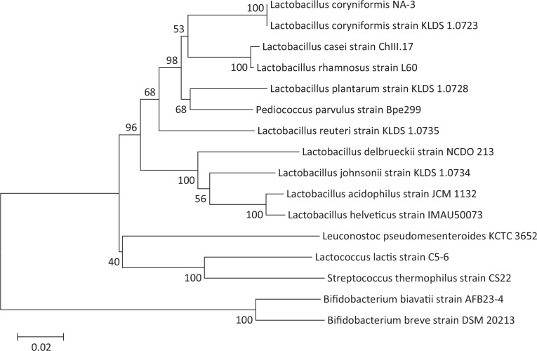
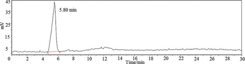
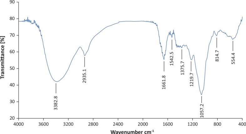
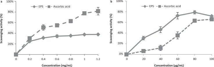
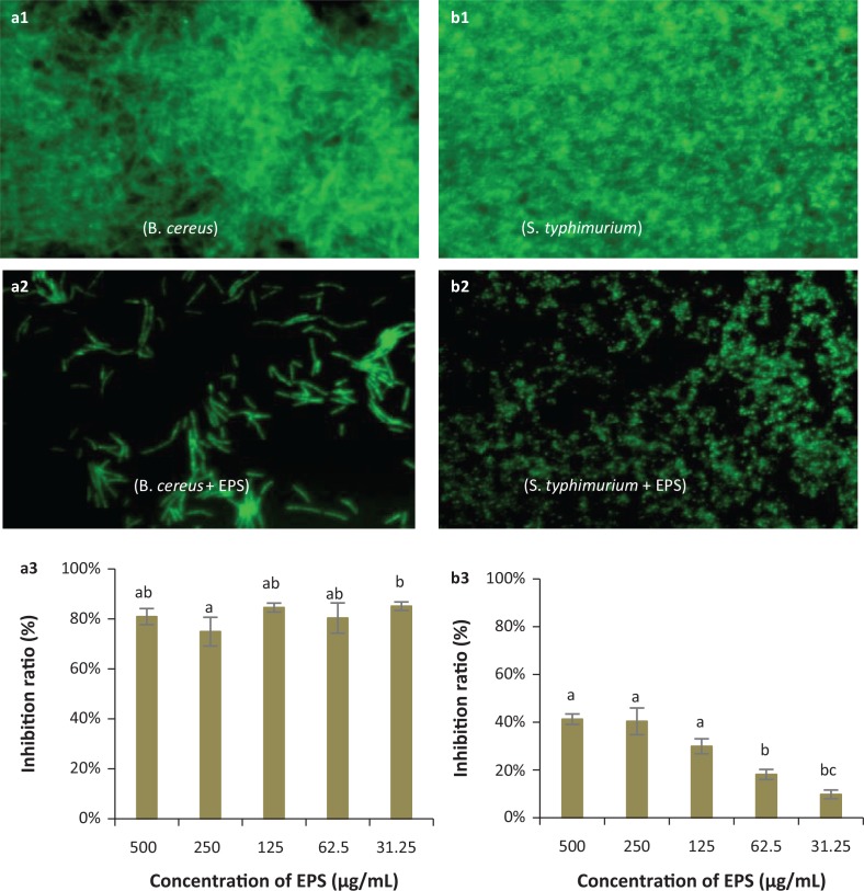
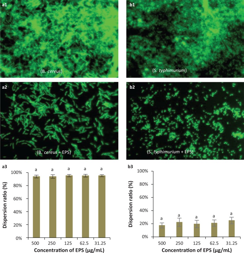
Methods: Lactobacillus coryniformis NA-3 was identified with 16S rDNA amplification and Neighbor Joining (NJ) phylogenetic analysis. EPS derived from Lactobacillus coryniformis NA-3 (EPS-NA3) was analyzed, including compositions by high-performance liquid chromatography (HPLC), functional groups by Fourier-transform infrared spectroscopy (FT-IR) and glycosidic bond configuration by Hydrogen-1 Nuclear Magnetic Resonance (1H NMR). Antioxidant activity of EPS was evaluated with hydroxyl and superoxide radical-scavenging. Anti-biofilm activities of EPS-NA3 were checked through inhibition and dispersion.

Results: The monosaccharide composition of EPS included α-rhamnose, α-mannose, α-galactose, and α-glucose in a ratio of 2.6:1.0:5.0:3.3. The free radical-scavenging abilities of EPS-NA3 were 37.77% ± 1.56% and 78.87% ± 3.07% on hydroxyl and superoxide reactive oxygen species respectively. Moreover, EPS-NA3 attenuated the formation of Bacillus cereus and Salmonella typhimurium biofilms by inhibition ratios of approximately 80% and 40% respectively. Additionally, treatment with EPS-NA3 dispersed established biofilms of B. cereus and S. typhimurium by approximately 90% and 20% respectively.

Conclusion: These results suggest that EPS-NA3 may be developed as antioxidant and anti-biofilm agents for industrial and clinical applications due to its capacity of scavenging free radicals, inhibition of bacterial biofilm formation, and dispersion of established biofilms.

Keywords: Lactobacillus coryniformis NA-3; anti-biofilm; antioxidant; dispersion; exopolysaccharide; inhibition.


Conflict of interest statement

The authors declare no potential conflicts of interest.

Figures

Fig. 1
Fig. 1
Neighbor joining-dendrogram showing the phylogenetic relationship between16S rDNA nucleotide sequences of Lactobacillus spp., obtained from the GenBank database, and Lactobacillus coryniformis NA-3 16S sequence, isolated in this study.
Fig. 2
Fig. 2
HPLC chromatogram showed the molecular weight of the EPS produced by Lactobacillus coryniformis NA-3, calculated by linear regression with dextran standards.
Fig. 3
Fig. 3
The FT-IR spectrum of exopolysaccharide (EPS) produced by Lactobacillus coryniformis NA-3 with the KBr pellet pressing method; x-axis is wave number (cm−1) and y-axis is transmittance (%).
Fig. 4
Fig. 4
The1H NMR spectrum of EPS-NA3.
Fig. 5
Fig. 5
Free radical-scavenging activities of EPS derived from Lactobacillus coryniformis NA-3 and ascorbic acid (positive control): hydroxyl radicals (a) and superoxide radicals (b). Data are represented as mean ± standard deviation (SD) of three replicates per experiment. Three independent experiments were conducted for each assay.
Fig. 6
Fig. 6
Representative micrographs (40×) showing the inhibitory effects of EPS on biofilm formation by pathogenic bacteria: Bacillus cereus (a1, a2, a3) and Salmonella typhimurium (b1, b2, b3) and spectrophotometric analyses of EPS biofilm inhibition. (a1, b1): Negative controls (pathogen only, no EPS) for biofilm formation by fluorescence microscopy; (a2, b2): Biofilm formation in the presence of EPS observed by fluorescence microscopy; (a3, b3): Inhibition ratios of EPS against B. cereus and S. typhimurium biofilms respectively. a,b,cDifferent letters indicate significant differences between concentrations (P < 0.05).
Fig. 7
Fig. 7
Representative micrographs (40×) showing the dispersion activity by EPS against established biofilms of the pathogenic bacteria Bacillus cereus (a1, a2, a3) and Salmonella typhimurium (b1, b2, b3), and spectrophotometric analyses of EPS-mediated dispersion of biofilms produced by these species. (a1, b1): Observation by fluorescence microscopy of negative controls (pathogens only, no EPS) for biofilm dispersion; (a2, b2): Observation by fluorescence microscopy of biofilm dispersion by Lactobacillus coryniformis NA-3-derived EPS; (a3, b3): Dispersion ratios of EPS on different bacterial biofilms. a,b,cDifferent letters indicate significant differences between treatments (P < 0.05).

References

    1. Fontana C, Li S, Yang Z, Widmalm G. Structural studies of the exopolysaccharide from Lactobacillus plantarum C88 using NMR spectroscopy and the program CASPER. Carbohyd Res 2015; 402: 87–94. doi: 10.1016/j.carres.2014.09.003 - DOI - PubMed
    1. Jia K, Tao X, Liu Z, Zhan H, He W, Zhang Z, et al. . Characterization of novel exopolysaccharide of Enterococcus faecium WEFA23 from infant and demonstration of its in vitro biological properties. Int J Biol Macromol 2019; 128: 710–17. doi: 10.1016/j.ijbiomac.2018.12.245 - DOI - PubMed
    1. Ye G, Chen Y, Wang C, Yang R, Bin X. Purification and characterization of exopolysaccharide produced by Weissellacibaria YB-1 from pickle Chinese cabbage. Int J Biol Macromol 2018; 120: 1315–21. doi: 10.1016/j.ijbiomac.2018.09.019 - DOI - PubMed
    1. Behare PV, Singh R, Kumar M, Prajapati JB, Singh RP. Exopolysaccharides of lactic acid bacteria: a review. J Food Sci Tech Mys 2009; 46(1): 1–11. doi: 10.1111/j.1750-3841.2008.01020.x - DOI
    1. Xu Y, Cui Y, Yue F, Liu L, Shan Y, Liu B, et al. . Exopolysaccharides produced by lactic acid bacteria and bifido bacteria: structures, physiochemical functions and applications in the food industry. Food Hydrocolloid 2019; 94: 475–499. doi: 10.1016/j.foodhyd.2019.03.032 - DOI