Review
. 2020 May 13:10:64.
doi: 10.1186/s13578-020-00426-y. eCollection 2020.

Synergies in exosomes and autophagy pathways for cellular homeostasis and metastasis of tumor cells

Affiliations
Review

Synergies in exosomes and autophagy pathways for cellular homeostasis and metastasis of tumor cells

Leila Salimi et al. Cell Biosci. .

Abstract

Background: Eukaryotic cells demonstrate two tightly linked vesicular transport systems, comprising intracellular vesicle transport and extracellular vesicle transport system. Intracellular transport vesicles can translocate biomolecules between compartments inside the cell, for example, proteins from the rough endoplasmic reticulum to the Golgi apparatus. Whereas, the secreted vesicles so-called extracellular vesicles facilitate the transport of biomolecules, for example, nucleic acids, proteins and lipids between cells. Vesicles can be formed during the process of endocytosis or/and autophagy and not only act as mediators of intra- and inter-cellular communication but also represent pathological conditions of cells or tissues.

Methods: In this review, we searched articles in PubMed, published between 2000 and 2020, with following terms: autophagy, autophagocytosis, transport vesicles, lysosomes, endosomes, exocytosis, exosomes, alone or in different combinations. The biological functions that were selected based on relevancy to our topic include cellular homeostasis and tumorigenesis.

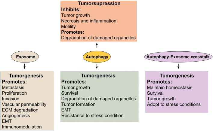
Results: The searched literature shows that there is a high degree of synergies between exosome biogenesis and autophagy, which encompass endocytosis and endosomes, lysosomes, exocytosis and exosomes, autophagocytosis, autophagosomes and amphisomes. These transport systems not only maintain cellular homeostasis but also operate synergically against fluctuations in the external and internal environment such as during tumorigenesis and metastasis. Additionally, exosomal and autophagic proteins may serve as cancer diagnosis approaches.

Conclusion: Exosomal and autophagy pathways play pivotal roles in homeostasis and metastasis of tumor cells. Understanding the crosstalk between endomembrane organelles and vesicular trafficking may expand our insight into cooperative functions of exosomal and autophagy pathways during disease progression and may help to develop effective therapies against lysosomal diseases including cancers and beyond.

Keywords: Autophagosomes; Autophagy; Autophagy associated tumorigenesis; Autophagy-mediated exosomes; Cancer cell metastasis; Endosomes; Extracellular vesicles.


Conflict of interest statement

Competing interestsThere is no any conflict of interest by the authors competing interests.

Figures

Fig. 1
Fig. 1
A schematic illustration of three types of autophagy and key regulatory molecules of autophagy flux inside cell. a: Three types of autophagy may occur in cell; microautophagy, chaperone-mediated autophagy, and macroautophagy [19, 20]. Microautophagy is the process during which damaged biomolecules are directly sorted into lysosomes. In chaperone-mediated autophagy, HSC70 identifies proteins containing specific motifs (KFERQ) and sorts them into lysosome through interaction with LAMP2A molecules placed on lysosome membrane. Macroautophagy mediates the lysosomal degradation of damaged proteins and organelles through 4 steps including initiation, nucleation, maturation, and finally fusion the autophagosome with lysosomes. Several proteins such as ULK, ATG13, FIP200, ARG101, Beclin-1, ATG14L, ATG5, ATG12, ATG16L, LC3, and PE, in different steps, mediate the formation of autophagosome [19, 20]. b: Once autophagy is induced, cytoplasmic dysfunctional molecules are encapsulated via double membranes, beginning from the formation of the phagophore to the autophagosomes, which consequently fuse with lysosomes and then their cargo is degraded [24]. Several ATG-associated assemblies including ULK-1 initiation complex, the PI3K III nucleation complex, the ATG12 conjugated complex, and the LC3 conjugation complex are involved in autophagy flux, which finally direct cytoplasmic dysfunctional molecules into lysosomes [24]. Stress condition such as starvation, energy depletion, reactive oxygen species (ROS), and hypoxia inhibit mTOR and growth factors act as activators of mTOR. Inhibition of mTOR activates the ULK-1 initiation complex which, in turn, mediates initiation of autophagy flux. In this scenario, AGT9 and the PI3K III nucleation complex collaborate with the ULK-1 initiation complex and progress initiation step of autophagy [24]. These complexes are supported by the ATG12 conjugation complex and the LC3 conjugation complex for completing initiation step and formation of phagophore in nucleation step. In order to formation of the ATG12 conjugation complex, ATG12 attaches to ATG5 and ATG16L1, and then the PI3P-binding complex (WIPIs and DFCP1) joins them to form the ATG12 conjugation complex. Formation of the ATG12 conjugation complex then facilitates connection of LC3 conjugation complex to newly formed phagophore in nucleation step, at this moment, ATG4 catalyzes the formation of LC3-I from LC3. Next, conjugation of PE with LC3-I, in presence of ATG7 and ATG3, forms LC3-II. This molecule is assimilated into phagophore and autophagosomal membranes, where LC3-II interacts with cargo receptors, which harbor LIRs [24]. DFCP1, zinc-finger; ECM, extracellular matrix; FYVE domain-containing protein 1; LC3, microtubule-associated protein light chain 3; LIRs, LC3-interacting motifs; PE: phosphatidylethanolamine ULK-1, Unc-51-like kinase 1; WIPIs, WD repeat domain phosphoinositide-interacting proteins
Fig. 2
Fig. 2
Formation of exosomes inside cell. Exosomes are nano-sized vesicles generated from endocytic pathway [5]. They are formed from inward budding of the membrane of multivesicular bodies (MVBs), late endosomes of endocytic pathway, through ESCRT-dependent machinery which involves assortment of ubiquitinated cargo. In addition, different proteins and lipids including CD63 and ceramides mediate exosome biogenesis which known as ESCRT-independent machinery [5, 38]. MVB’s cargo is provided with different sorting molecules located on MVB’s membrane, cytoplasm, and Golgi apparatus. Different Rab-GTPases such as Rab7, Rab11, Rab27, and Rap35 preferentially mediate intracellular trafficking of MVBs. MBVs may back fuse to the plasma membrane and recycle biomolecules to the plasma membrane or present specific biomolecules (such as major histocompatibility complex (MHC) proteins) (1). SNARE and Rab-GTPase (Rab11, Rab27, and Rab35) proteins facilitate the fusion of MVBs with the plasma membrane in order to release exosomes into extracellular environment (2). In degradation pathway, MVBs can fuse with lysosomes for hydrolyzing their cargo (3). EE, early endosome; ER, endoplasmic reticulum; GA, Golgi apparatus; N, nucleus
Fig. 3
Fig. 3
Crosstalk between exosome biogenesis and autophagy. Link between exosome biogenesis and autophagy pathways exists not only at molecular level but also at membranous vesicles such as amphisomes. In this cooperative action, various Rab-GTPase proteins including Rab8a, Rab11, and Rab27 control the movement of vesicles between exosomal secretory pathway and autophagy at the cytoplasm. Autophagic proteins including LC3β, ATG5, and ATG16L1, on the MVB’s membrane, contribute to generate exosomes. Then the autophagic cargo can be secreted into extracellular milieu via exosomes. Additionally, the MVBs may fuse with autophagosome to make hybrid vesicles named amphisomes. Amphisomes cargo may be degraded by lysosomes or alternatively may fuse with the plasma membrane and secrete cargo into extracellular milieu. Amphisomes participate in packaging of annexin A2 (ANXA2) into exosomes; however, which cargo received from autophagosomes may sort into exosomes in amphisomes is still remains a mystery
Fig. 4
Fig. 4
A schematic diagram of key roles of exosomes, autophagy, and autophagy-exosomes crosstalk in cancer metastasis

References

    1. Conner SD, Schmid SL. Regulated portals of entry into the cell. Nature. 2003;422(6927):37–44. doi: 10.1038/nature01451. - DOI - PubMed
    1. Tokarev AA, Alfonso A, Segev N. Overview of intracellular compartments and trafficking pathways. Trafficking Inside Cells: Springer; 2009. pp. 3–14.
    1. Bareford LM, Swaan PW. Endocytic mechanisms for targeted drug delivery. Adv Drug Deliv Rev. 2007;59(8):748–758. doi: 10.1016/j.addr.2007.06.008. - DOI - PMC - PubMed
    1. Dupont N, Jiang S, Pilli M, Ornatowski W, Bhattacharya D, Deretic V. Autophagy-based unconventional secretory pathway for extracellular delivery of IL-1β. EMBO J. 2011;30(23):4701–4711. doi: 10.1038/emboj.2011.398. - DOI - PMC - PubMed
    1. Kowal J, Tkach M, Théry C. Biogenesis and secretion of exosomes. Curr Opin Cell Biol. 2014;29:116–125. doi: 10.1016/j.ceb.2014.05.004. - DOI - PubMed

LinkOut - more resources