. 2020 Jun 2;31(9):107725.
doi: 10.1016/j.celrep.2020.107725. Epub 2020 May 18.

Cross-reactive Antibody Response between SARS-CoV-2 and SARS-CoV Infections

Affiliations

Cross-reactive Antibody Response between SARS-CoV-2 and SARS-CoV Infections

Huibin Lv et al. Cell Rep. .

Abstract

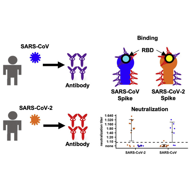
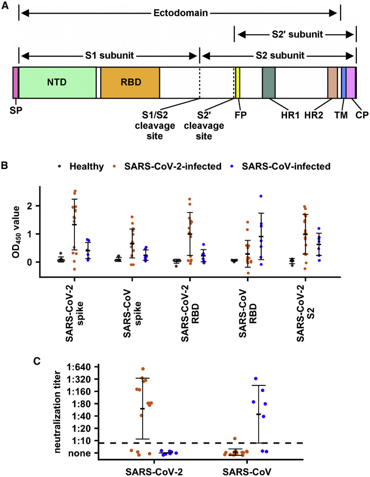
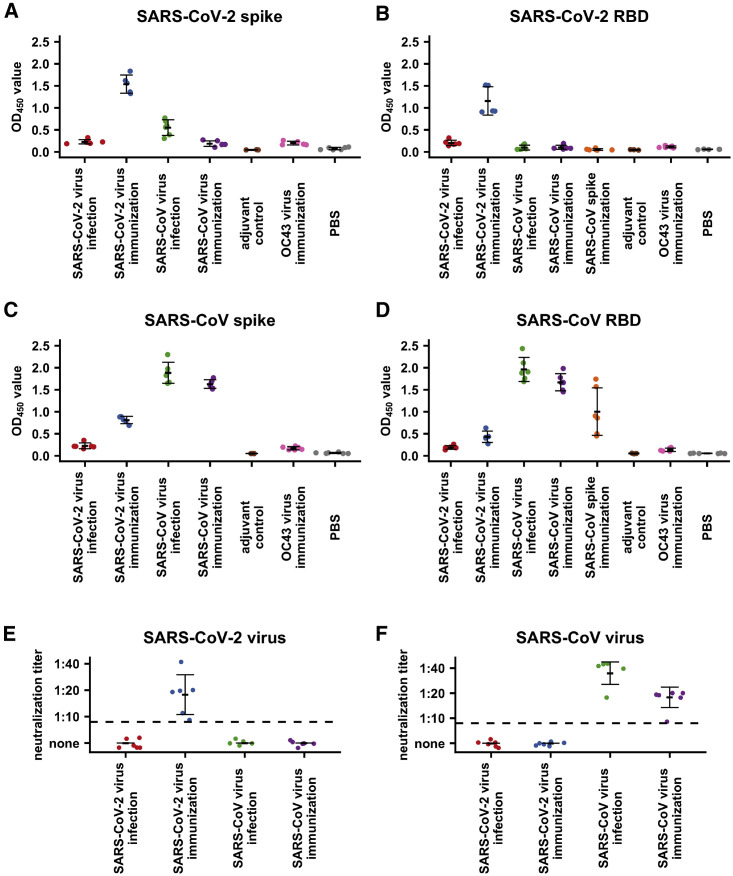
The World Health Organization has declared the ongoing outbreak of COVID-19, which is caused by a novel coronavirus SARS-CoV-2, a pandemic. There is currently a lack of knowledge about the antibody response elicited from SARS-CoV-2 infection. One major immunological question concerns antigenic differences between SARS-CoV-2 and SARS-CoV. We address this question by analyzing plasma from patients infected by SARS-CoV-2 or SARS-CoV and from infected or immunized mice. Our results show that, although cross-reactivity in antibody binding to the spike protein is common, cross-neutralization of the live viruses may be rare, indicating the presence of a non-neutralizing antibody response to conserved epitopes in the spike. Whether such low or non-neutralizing antibody response leads to antibody-dependent disease enhancement needs to be addressed in the future. Overall, this study not only addresses a fundamental question regarding antigenicity differences between SARS-CoV-2 and SARS-CoV but also has implications for immunogen design and vaccine development.


Conflict of interest statement

Declaration of Interests The authors declare no competing interests.

Figures

None
Graphical abstract
Figure 1
Figure 1
Human Serological Responses to SARS-CoV-2 (A) Schematic diagram of the SARS-CoV-2 spike protein. Locations of secretion signal peptide (SP), N-terminal domain (NTD), receptor-binding domain (RBD), S1/S2 cleavage site, S2′ cleavage site, fusion peptide (FP), heptad repeat 1 (HR1), heptad repeat 2 (HR2), transmembrane domain (TM), and cytoplasmic domain (CP) are indicated. Regions corresponding to the S1, S2, S2′ subunits, and ectodomain are also indicated. (B and C) Binding of plasma from healthy donors and SARS-CoV-2-infected patients to SARS-CoV-2 spike protein, SARS-CoV-2 RBD protein, SARS-CoV-2 S2 subunit, SARS-CoV spike protein, and SARS-CoV RBD protein were measured by ELISA (B). The mean OD450 values calculated after testing each plasma sample in triplicate are shown. (C) Neutralization activities of plasma from SARS-CoV-2-infected patients to SARS-CoV-2 and SARS-CoV viruses were measured. Dashed line represents the lower detection limit. Black lines indicate means ± SD. (B and C) Grey, plasma samples from healthy donors; orange, plasma samples from SARS-CoV-2-infected patients; blue, plasma samples from SARS-CoV-infected patients. The value from each dot in the figure was taken by the means of two replicates in the same assay.
Figure 2
Figure 2
Mouse Serological Response to SARS-CoV-2 and SARS-CoV (A–D) Binding of plasma from OC43-CoV-immunized mice, SARS-CoV-immunized mice, SARS-CoV-infected mice, and mock-immunized mice against (A) SARS-CoV-2 spike protein, (B) SARS-CoV-2 RBD protein, (C) SARS-CoV spike protein, and (D) SARS-CoV RBD protein were measured by ELISA. Because both SARS-CoV spike protein and SARS-CoV-2 spike contained a C-terminal foldon domain, binding of plasma from mice immunized with the SARS-CoV spike protein plasma was not tested against spike proteins from SARS-CoV and SARS-CoV-2. The mean OD450 values calculated after testing each plasma sample in triplicate are shown. (E and F) Neutralization activities of plasma from mice infected or immunized by SARS-CoV-2 or SARS-CoV to (E) SARS-CoV-2 virus or (F) SARS-CoV virus were measured. Dashed line represents the lower detection limit. Black lines indicate means ± SD. The value from each dot in the figure was taken by the means of two replicates in the same assay.

Update of

References

    1. Bajic G., Maron M.J., Adachi Y., Onodera T., McCarthy K.R., McGee C.E., Sempowski G.D., Takahashi Y., Kelsoe G., Kuraoka M., Schmidt A.G. Influenza antigen engineering focuses immune responses to a subdominant but broadly protective viral epitope. Cell Host Microbe. 2019;25:827–835.e6. - PMC - PubMed
    1. Bangaru S., Lang S., Schotsaert M., Vanderven H.A., Zhu X., Kose N., Bombardi R., Finn J.A., Kent S.J., Gilchuk P., et al. A site of vulnerability on the influenza virus hemagglutinin head domain trimer interface. Cell. 2019;177:1136–1152.e18. - PMC - PubMed
    1. Bao L., Deng W., Huang B., Gao H., Liu J., Ren L., Wei Q., Yu P., Xu Y., Qi F., et al. The pathogenicity of SARS-CoV-2 in hACE2 transgenic mice. Nature. 2020 doi: 10.1101/2020.02.07.939389. - DOI - PubMed
    1. Bootz A., Karbach A., Spindler J., Kropff B., Reuter N., Sticht H., Winkler T.H., Britt W.J., Mach M. Protective capacity of neutralizing and non-neutralizing antibodies against glycoprotein B of cytomegalovirus. PLoS Pathog. 2017;13:e1006601. - PMC - PubMed
    1. Burke C.W., Froude J.W., Miethe S., Hülseweh B., Hust M., Glass P.J. Human-like neutralizing antibodies protect mice from aerosol exposure with western equine encephalitis virus. Viruses. 2018;10:147. - PMC - PubMed

Publication types

MeSH terms

LinkOut - more resources