Review
. 2020 Apr 23:7:77-89.
doi: 10.2147/JHC.S224471. eCollection 2020.

Angiogenesis in Hepatocellular Carcinoma; Pathophysiology, Targeted Therapy, and Role of Imaging

Affiliations
Review

Angiogenesis in Hepatocellular Carcinoma; Pathophysiology, Targeted Therapy, and Role of Imaging

Ahmed W Moawad et al. J Hepatocell Carcinoma. .

Abstract

Hepatocellular carcinoma (HCC) is one of the most common tumors worldwide, usually occurring on a background of liver cirrhosis. HCC is a highly vascular tumor in which angiogenesis plays a major role in tumor growth and spread. Tumor-induced angiogenesis is usually related to a complex interplay between multiple factors and pathways, with vascular endothelial growth factor being a major player in angiogenesis. In the past decade, understanding of tumor-induced angiogenesis has led to the emergence of novel anti-angiogenic therapies, which act by reducing neo-angiogenesis, and improving patient survival. Currently, Sorafenib and Lenvatinib are being used as the first-line treatment for advanced unresectable HCC. However, a disadvantage of these agents is the presence of numerous side effects. A major challenge in the management of HCC patients being treated with anti-angiogenic therapy is effective monitoring of treatment response, which decides whether to continue treatment or to seek second-line treatment. Several criteria can be used to assess response to treatment, such as quantitative perfusion on cross-sectional imaging and novel/emerging MRI techniques, including a host of known and emerging biomarkers and radiogenomics. This review addresses the pathophysiology of angiogenesis in HCC, accurate imaging assessment of angiogenesis, monitoring effects of anti-angiogenic therapy to guide future treatment and assessing prognosis.

Keywords: Sorafenib; angiogenesis; anti-angiogenic therapy; hepatocellular carcinoma.


Conflict of interest statement

The authors report no conflicts of interest in this work.

Figures
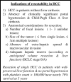
Figure 1
Figure 1
Indication of HCC resection as stated by ASSLD guidelines for treatment HCC 2018. Data from Poon et al.
Figure 2
Figure 2
Contrast-enhanced axial CT images in a patient with HCC. (A) Demonstrates a large dominant mass within segment IV. (B) Acquired 6 months after Sorafenib therapy demonstrates significant reduction in the vascularity of the tumor with no significant relative decrease in size. Note the shrunken liver (arrowhead) with worsening cirrhosis and ascites (arrow).
Figure 3
Figure 3
Axial CT images of the liver with advanced HCC: pretreatment (A) and post-treatment with Sorafenib (2 months interval (B) and 4 months interval (C)). Lesions demonstrate slightly increased size but decreased enhancement, suggesting treatment response.
Figure 4
Figure 4
Digital subtraction angiogram of trans-femoral injection of contrast through the hepatic artery revealed a focal hepatic lesion with progressive increased arterial uptake (“tumoral blush”), confirming that the main origin of the HCC supply is from the hepatic artery not portal vein, like the rest of the hepatic parenchyma.
Figure 5
Figure 5
Multiphasic CT scan of the liver with arterial phase (Panel (A), portal phase (B) and delayed phase (C) demonstrates the typical features of HCC with non-rim arterial hyper-enhancement, iso to hypoenhancing at the portal phase and washout of the contrast in the delayed phase. Note the capsular enhancement of the tumor on the delayed phase sequence (arrow).
Figure 6
Figure 6
Contrast enhanced MRI of abdomen (extracellular agent) shows a focal hepatic lesion in segment III (arrow) which is iso-intense to the surrounding liver parenchyma (no hyper-enhancement) in the arterial phase (A) and “washout” of the contrast in the delayed phase (B).
Figure 7
Figure 7
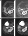
Axial MRI of the liver ((A1, A2) post contrast, (B1, B2) T2WI) showing segment VI HCC before and after treatment with Sorafenib. Notice in panel (A) post-treatment MRI (A2) shows HCC with decrease central enhancement (comparing to pre-treatment image (A1)) with geographical perilesional enhancement go with perfusion changes. Notice in image (B2) the increase in T2WI signal intensity (compared to (B1)) as a part of post-Sorafenib changes.
Figure 8
Figure 8
Multiphasic contrast enhanced MRI of liver with use of Gadoxetate sodium as contrast agent (hepatobiliary). (A) Axial T2 weighted images fat-suppressed show left lobe focal lesion measuring 10 cm showing high T2 signal intensity. (B) Axial T1 weighted images (T1WI) without contrast showing heterogeneous low signal intensity in the lesion. Axial T1WI with IV Gadoxetate sodium in the arterial phase showing non rim hyper-enhancement (C) and hepatobiliary phase (D) (3D dynamic protocol with 20 min delayed) showing the classic “washout” of the contrast with relatively lower signal than the surrounding liver parenchyma. (E, F) axial DWI (b-value = 400 and 800 s/mm, respectively) showing heterogonous restricted diffusion.

References

    1. Mohammadian M, Mahdavifar N, Mohammadian-hafshejani A. Liver cancer in the world: epidemiology, incidence, mortality and risk factors. WCRJ. 2018;5(2):e1082.
    1. Mittal S, El-serag HB. Epidemiology of hepatocellular carcinoma: consider the population. J Clin Gastroenterol. 2013;47(Suppl(0)):S2–S6. doi:10.1097/MCG.0b013e3182872f29 - DOI - PMC - PubMed
    1. Llovet JM, Burroughs A, Bruix J. Hepatocellular carcinoma. Lancet. 2003;362(9399):1907–1917. doi:10.1016/S0140-6736(03)14964-1 - DOI - PubMed
    1. Seeff LB. Introduction: the burden of hepatocellular carcinoma. Gastroenterology. 2004;127(5,Supplement 1):S1–S4. doi:10.1053/j.gastro.2004.09.010 - DOI - PubMed
    1. Janevska D, Chaloska-ivanova V, Janevski V. Hepatocellular carcinoma: risk factors, diagnosis and treatment. Open Access Maced J Med Sci. 2015;3(4):732–736. doi:10.3889/oamjms.2015.111 - DOI - PMC - PubMed