. 2020 Apr 30:8:373.
doi: 10.3389/fbioe.2020.00373. eCollection 2020.

Stiffness Matters: Fine-Tuned Hydrogel Elasticity Alters Chondrogenic Redifferentiation

Affiliations

Stiffness Matters: Fine-Tuned Hydrogel Elasticity Alters Chondrogenic Redifferentiation

Barbara Bachmann et al. Front Bioeng Biotechnol. .

Abstract

Biomechanical cues such as shear stress, stretching, compression, and matrix elasticity are vital in the establishment of next generation physiological in vitro tissue models. Matrix elasticity, for instance, is known to guide stem cell differentiation, influence healing processes and modulate extracellular matrix (ECM) deposition needed for tissue development and maintenance. To better understand the biomechanical effect of matrix elasticity on the formation of articular cartilage analogs in vitro, this study aims at assessing the redifferentiation capacity of primary human chondrocytes in three different hydrogel matrices of predefined matrix elasticities. The hydrogel elasticities were chosen to represent a broad spectrum of tissue stiffness ranging from very soft tissues with a Young's modulus of 1 kPa up to elasticities of 30 kPa, representative of the perichondral-space. In addition, the interplay of matrix elasticity and transforming growth factor beta-3 (TGF-β3) on the redifferentiation of primary human articular chondrocytes was studied by analyzing both qualitative (viability, morphology, histology) and quantitative (RT-qPCR, sGAG, DNA) parameters, crucial to the chondrotypic phenotype. Results show that fibrin hydrogels of 30 kPa Young's modulus best guide chondrocyte redifferentiation resulting in a native-like morphology as well as induces the synthesis of physiologic ECM constituents such as glycosaminoglycans (sGAG) and collagen type II. This comprehensive study sheds light onto the mechanobiological impact of matrix elasticity on formation and maintenance of articular cartilage and thus represents a major step toward meeting the need for advanced in vitro tissue models to study both re- and degeneration of articular cartilage.

Keywords: 3D cell culture; Young’s modulus; cartilage; chondrocytes; extracellular matrix; hydrogel.


Figures

FIGURE 1
FIGURE 1
Overview of workflow for redifferentiation analysis of primary human chondrocytes in hydrogels of different elasticities. After rheological measurements for obtaining hydrogels of defined elasticities of 1 kPa, 15 kPa, and 30 kPa Youngs’s modulus, primary human chondrocytes where embedded and cultivated for 3 weeks. Chondrogenic redifferentiation was subsequently assessed by viability, histology and quantitative analysis of cartilage matrix components by GAG Assay and RT-qPCR.
FIGURE 2
FIGURE 2
Viability of primary chondrocytes in fibrin, silk fibrin and PEG-dextran hydrogels depicted as mean with SD (n = 9). Fibrin and silk/fibrin hydrogels display high viabilities irrespective of stiffness while viability in PEG-dextran hydrogels declines rapidly with increasing elasticity. Epifluorescent exemplary images of Live(Calcein-AM)/Dead staining (Ethidium-H1) in 15 kPa hydrogels without growth factor addition (below). Scale bar 100 μm.
FIGURE 3
FIGURE 3
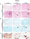
Histological images of 4 μm thick sections of primary chondrocytes in fibrin hydrogels of 1 kPa, 15 kPa, and 30 kPa elasticity stained with (A) H&E for morphology, (B) alcian blue for sGAG and (C) collagen II. Scale bar 50 μm. Arrows highlight visible morphological differences in the presence of increasing matrix elasticity and growth factor stimulation, where elongated, fibroblastic morphology is found in softer hydrogels, and chondrotypic spherical morphology in 30 kPa hydrogels. Growth factor stimulation resulted in an overall increase in cell numbers with stiffness-depended cell distribution including cellular cluster formations in 30 kPa hydrogels.
FIGURE 4
FIGURE 4
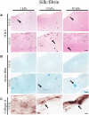
Histological images of 4 μm thick sections primary chondrocytes in silk/fibrin hydrogels of 1 kPa, 15 kPa and 30 kPa elasticity stained with (A) H&E for morphology, (B) alcian blue for sGAG and (C) collagen II. Scale bar 50 μm. Arrows highlight visible in the presence of increasing matrix elasticity and growth factor stimulation, resulting in enhanced chondrotypic sphericity with increasing hydrogel stiffness and cell cluster formation at 30 kPa hydrogels. Additional stimulation with growth factor resulted in massive formation of cartilage matrix collagen II around the clusters.
FIGURE 5
FIGURE 5
(A) Gene expression of redifferentiation indices and aggrecan expression in the presence of increasing hydrogel stiffness, resulting in incremental increases of collagen type II and aggrecan synthesis using fibrin hydrogels. In turn, in the presence of PEG-dextran hydrogels native amount of aggrecan synthesis was observed (results should be interpreted with caution due to low cell vitality). (B) Quantification of synthesized sGAG per μg DNA in fibrin, silk/fibrin and PEG-dextran hydrogel of varied elasticity again showed an improved redifferentiation behavior in fibrin hydrogels with increasing matrix elasticity. All results depicted as mean with SEM. n = 9; *p < 0.1; **p < 0.05; ***p < 0.01; ****p < 0.001.

References

    1. Allen J. L., Cooke M. E., Alliston T. (2012). ECM stiffness primes the TGFbeta pathway to promote chondrocyte differentiation. Mol. Biol. Cell. 23 3731–3742. 10.1091/mbc.e12-03-0172 - DOI - PMC - PubMed
    1. Camarero-Espinosa S., Rothen-Rutishauser B., Foster E. J., Weder C. (2016). Articular cartilage: from formation to tissue engineering. Biomater. Sci. 4 734–767. 10.1039/c6bm00068a - DOI - PubMed
    1. Caron M. M., Emans P. J., Coolsen M. M., Voss L., Surtel D. A., Cremers A., et al. (2012). Redifferentiation of dedifferentiated human articular chondrocytes: comparison of 2D and 3D cultures. Osteoarthritis Cartilage 20 1170–1178. 10.1016/j.joca.2012.06.016 - DOI - PubMed
    1. Carter D. R., Beaupre G. S., Wong M., Smith R. L., Andriacchi T. P., Schurman D. J. (2004). The mechanobiology of articular cartilage development and degeneration. Clin. Orthop. Relat. Res. 427 S69–S77. - PubMed
    1. Discher D. E., Mooney D. J., Zandstra P. W. (2009). Growth factors, matrices, and forces combine and control stem cells. Science 324 1673–1677. 10.1126/science.1171643 - DOI - PMC - PubMed

LinkOut - more resources