

Femoral Tunnel Placement Analysis in ACL Reconstruction Through Use of a Novel 3-Dimensional Reference With Biplanar Stereoradiographic Imaging
- PMID: 32426408
- PMCID: PMC7219010
- DOI: 10.1177/2325967120915709
Femoral Tunnel Placement Analysis in ACL Reconstruction Through Use of a Novel 3-Dimensional Reference With Biplanar Stereoradiographic Imaging
Abstract
Background: The femoral-sided anatomic footprint of the anterior cruciate ligament (ACL) has been widely studied during the past decades. Nonanatomic placement is an important cause of ACL reconstruction (ACLR) failure.
Purpose: To describe femoral tunnel placement in ACLR through use of a comprehensive 3-dimensional (3D) cylindrical coordinate system combining both the traditional clockface technique and the quadrant method. Our objective was to validate this technique and evaluate its reproducibility.
Study design: Descriptive laboratory study.
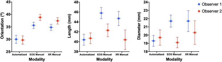
Methods: The EOS Imaging System was used to make 3D models of the knee for 37 patients who had undergone ACLR. We designed an automated cylindrical reference software program individualized to the distal femoral morphology of each patient. Cylinder parameters were collected from 2 observers' series of 3D models. Each independent observer also manually measured the corresponding parameters using a lateral view of the 3D contours and a 2-dimensional stereoradiographic image for the corresponding patient.
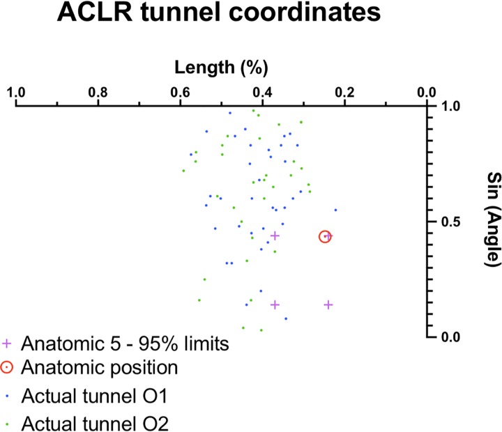
Results: The average cylinder produced from the first observer's EOS 3D models had a 30.0° orientation (95% CI, 28.4°-31.5°), 40.4 mm length (95% CI, 39.3-41.4 mm), and 19.3 mm diameter (95% CI, 18.6-20.0 mm). For the second observer, these measurements were 29.7° (95% CI, 28.1°-31.3°), 40.7 mm (95% CI, 39.7-41.8 mm), and 19.7 mm (95% CI, 18.8-20.6 mm), respectively. Our method showed moderate intertest intraclass correlation among all 3 measuring techniques for both length (r = 0.68) and diameter (r = 0.63) but poor correlation for orientation (r = 0.44). In terms of interobserver reproducibility of the automated EOS 3D method, similar results were obtained: moderate to excellent correlations for length (r = 0.95; P < .001) and diameter (r = 0.66; P < .001) but poor correlation for orientation (r = 0.29; P < .08). With this reference system, we were able to describe the placement of each individual femoral tunnel aperture, averaging a difference of less than 10 mm from the historical anatomic description by Bernard et al.
Conclusion: This novel 3D cylindrical coordinate system using biplanar, stereoradiographic, low-irradiation imaging showed a precision comparable with standard manual measurements for ACLR femoral tunnel placement. Our results also suggest that automated cylinders issued from EOS 3D models show adequate accuracy and reproducibility.
Clinical relevance: This technique will open multiple possibilities in ACLR femoral tunnel placement in terms of preoperative planning, postoperative feedback, and even intraoperative guidance with augmented reality.
Keywords: 3D modeling; ACL; knee; stereoradiographic imaging.
© The Author(s) 2020.
Conflict of interest statement
One or more of the authors has declared the following potential conflict of interest or source of funding: This study was sponsored by grants from the Natural Sciences and Engineering Research Council of Canada, Canada Research Chairs, Mitacs, and MEDTEQ. T.C. and J.A.D.G. have received research support and royalties from EOS Imaging and have licensed patents to EOS Imaging. AOSSM checks author disclosures against the Open Payments Database (OPD). AOSSM has not conducted an independent investigation on the OPD and disclaims any liability or responsibility relating thereto.
Figures


References
-
- Behrend H, Stutz G, Kessler MA, Rukavina A, Giesinger K, Kuster MS. Tunnel placement in anterior cruciate ligament (ACL) reconstruction: quality control in a teaching hospital. Knee Surg Sports Traumatol Arthrosc. 2006;14(11):1159–1165. - PubMed
-
- Bernard M, Hertel P, Hornung H, Cierpinski T. Femoral insertion of the ACL: radiographic quadrant method. Am J Knee Surg. 1997;10(1):14–21. - PubMed
-
- Burkart A, Debski RE, McMahon PJ, et al. Precision of ACL tunnel placement using traditional and robotic techniques. Comput Aided Surg. 2001;6(5):270–278. - PubMed
-
- Cicchetti DV. Guidelines, criteria, and rules of thumb for evaluating normed and standardized assessment instruments in psychology. Psychol Assess. 1994;6(4):284–290.
-
- Colombet P, Robinson J, Christel P, et al. Morphology of anterior cruciate ligament attachments for anatomic reconstruction: a cadaveric dissection and radiographic study. Arthroscopy. 2006;22(9):984–992. - PubMed
LinkOut - more resources
Full Text Sources
Miscellaneous
