. 2020 Apr 17;6(16):eaaz4316.
doi: 10.1126/sciadv.aaz4316. eCollection 2020 Apr.

Nanoparticle elasticity regulates phagocytosis and cancer cell uptake

Affiliations

Nanoparticle elasticity regulates phagocytosis and cancer cell uptake

Yue Hui et al. Sci Adv. .

Abstract

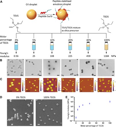
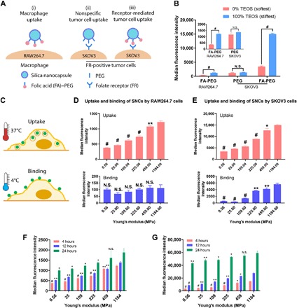
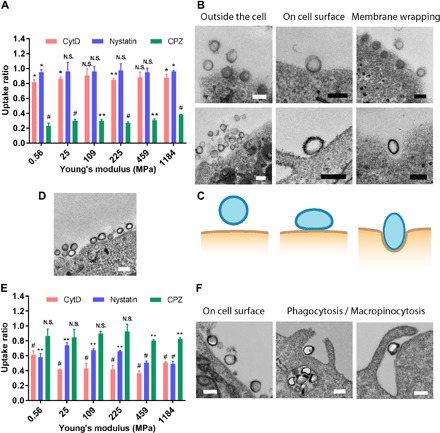
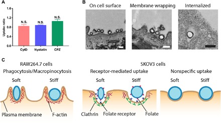
The ability of cells to sense external mechanical cues is essential for their adaptation to the surrounding microenvironment. However, how nanoparticle mechanical properties affect cell-nanoparticle interactions remains largely unknown. Here, we synthesized a library of silica nanocapsules (SNCs) with a wide range of elasticity (Young's modulus ranging from 560 kPa to 1.18 GPa), demonstrating the impact of SNC elasticity on SNC interactions with cells. Transmission electron microscopy revealed that the stiff SNCs remained spherical during cellular uptake. The soft SNCs, however, were deformed by forces originating from the specific ligand-receptor interaction and membrane wrapping, which reduced their cellular binding and endocytosis rate. This work demonstrates the crucial role of the elasticity of nanoparticles in modulating their macrophage uptake and receptor-mediated cancer cell uptake, which may shed light on the design of drug delivery vectors with higher efficiency.


Figures

Fig. 1
Fig. 1. Synthesis and characterization of SNCs.
(A) Schematic illustration of the synthesis of SNCs having controllable Young’s moduli. (B) TEM micrographs (scale bars, 200 nm) and (C) AFM height profiles (scale bars, 500 nm) of SNCs. (D) SEM micrographs (scale bars, 200 nm) of SNCs synthesized using 0% (softest) and 100% (stiffest) of TEOS. (E) Young’s moduli of SNCs (values are means ± SD, n = 15).
Fig. 2
Fig. 2. Effects of SNC elasticity on cellular interactions.
(A) Schematic illustration showing different types of cell-SNC interactions. (B) The uptake of the softest and the stiffest SNCs by RAW264.7 and SKOV3 cells; inset is the magnified graph of the first two groups. (C) The cellular uptake and binding of SNCs were measured at 4° and 37°C, respectively. (D) Four-hour cellular uptake and binding of FA-PEG–modified SNCs in RAW264.7 and (E) SKOV3 cells; values are compared to the SNCs having the highest Young’s modulus. (F) Four-, 12-, and 24-hour cellular uptake of FA-PEG–modified SNCs in RAW264.7 and (G) SKOV3 cells; at every time point, values are compared to the SNCs having the highest Young’s modulus. All values are means ± SD (n = 3, with *P < 0.05, **P < 0.01, and #P < 0.001; N.S., not significant).
Fig. 3
Fig. 3. Endocytic pathways and deformations of SNCs during cellular uptake.
(A) Uptake ratios of the FA-PEG–modified SNCs in SKOV3 cells. (B) TEM micrographs showing the morphological change of the stiffest (top) and softest (bottom) SNCs during receptor-mediated interactions with SKOV3 cells. (C) Schematic illustration showing the deformation of the softest SNCs during receptor-mediated cellular uptake. (D) Considerable proportion of SKOV3 cell surface can be covered by a large number of adhered SNCs. (E) Uptake ratios of the FA-PEG–modified SNCs in RAW264.7 cells. (F) TEM micrographs showing the morphological change of the softest SNCs during interactions with RAW264.7 cells. The uptake ratio represents the uptake of SNCs by cells treated with endocytic inhibitors normalized by the uptake by nontreated ones. Scale bars, 200 nm. All values are means ± SD (n = 3, with *P < 0.05, **P < 0.01, and #P < 0.001; N.S., not significant).
Fig. 4
Fig. 4. The endocytic pathway–dependent deformation of SNCs is a result of the associated proteins.
(A) Uptake ratios of the PEG-modified softest SNCs in SKOV3 cells. The uptake ratio represents the uptake of SNCs by cells treated with endocytic inhibitors normalized by the uptake by nontreated ones. (B) TEM micrographs showing the morphological change of the softest PEG-modified SNCs during interactions with SKOV3 cells; scale bars, 200 nm. (C) Schematic illustration showing the proteins associated in the three types of SNC-cell interactions. All values are means ± SD (n = 3).
Fig. 5
Fig. 5. The internalization kinetics of the softest (0% TEOS, 560 kPa) and the stiffest (100% TEOS, 1.18 GPa) SNCs in receptor-mediated uptake by SKOV3 cells.
(A) Fluorescence live-cell images showing the internalization of the softest and (B) stiffest FA-PEG–modified SNCs by SKOV3 cells and their transport to lysosomes over 60 min; scale bars, 20 μm. Representative time projection images showing the movement of the (C) softest and (D) stiffest SNCs during cellular uptake; tracks of the SNCs are shown in the magnified images of boxed regions. Yellow solid lines indicate the plasma membranes; scale bars, 10 μm. Instantaneous velocity of the (E) softest and (F) stiffest SNCs during stage I and stage II of cellular uptake. MSD analysis of the movement of the (G) softest and (H) stiffest SNCs during stage I and stage II of cellular uptake; insets are magnified graphs. (I) Time taken for the internalization of SNCs. Movements of the SNCs were only recorded for 15 min, as imaging longer than 15 min could lead to fluorescence quenching and focal plane shifting.

References

    1. Gillespie P. G., Walker R. G., Molecular basis of mechanosensory transduction. Nature 413, 194–202 (2001). - PubMed
    1. Luo T., Mohan K., Iglesias P. A., Robinson D. N., Molecular mechanisms of cellular mechanosensing. Nat. Mater. 12, 1064–1071 (2013). - PMC - PubMed
    1. Qiu Y., Brown A. C., Myers D. R., Sakurai Y., Mannino R. G., Tran R., Ahn B., Hardy E. T., Kee M. F., Kumar S., Bao G., Barker T. H., Lam W. A., Platelet mechanosensing of substrate stiffness during clot formation mediates adhesion, spreading, and activation. Proc. Natl. Acad. Sci. U.S.A. 111, 14430–14435 (2014). - PMC - PubMed
    1. Vogel V., Sheetz M., Local force and geometry sensing regulate cell functions. Nat. Rev. Mol. Cell Biol. 7, 265–275 (2006). - PubMed
    1. Discher D. E., Janmey P., Wang Y.-L., Tissue cells feel and respond to the stiffness of their substrate. Science 310, 1139–1143 (2005). - PubMed

Publication types

Substances