

Case Reports
doi: 10.1016/j.ekir.2020.05.005.
eCollection 2020 Jul.
Kidney Biopsy Findings in a Critically Ill COVID-19 Patient With Dialysis-Dependent Acute Kidney Injury: A Case Against "SARS-CoV-2 Nephropathy"
Affiliations
- PMID: 32426558
- PMCID: PMC7230145
- DOI: 10.1016/j.ekir.2020.05.005
Item in Clipboard
Case Reports
Kidney Biopsy Findings in a Critically Ill COVID-19 Patient With Dialysis-Dependent Acute Kidney Injury: A Case Against "SARS-CoV-2 Nephropathy"
Kidney Int Rep.
.
No abstract available
Figures
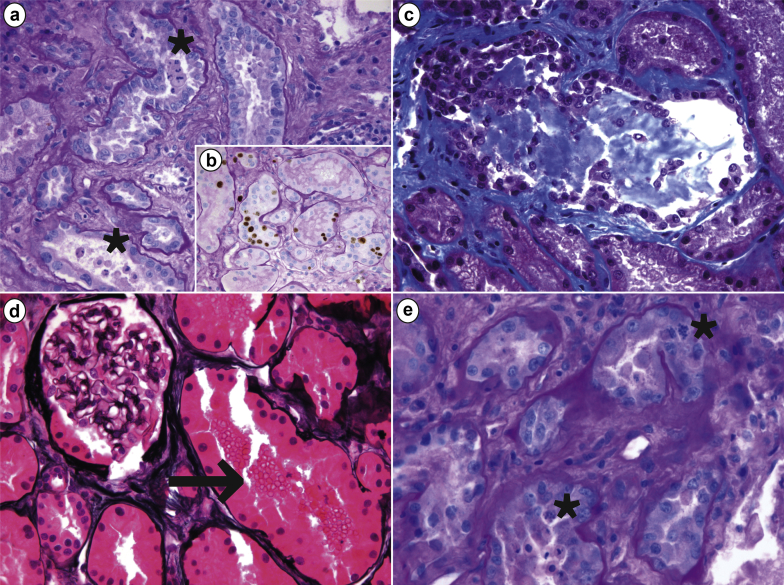
Morphologic features of acute tubular injury on renal biopsy of a critically ill patient with coronavirus disease 2019 (COVID-19) and acute kidney injury. (a) Diffuse tubular injury and focal tubular necrosis with several tubules showing intraluminal epithelial sloughing (asterisks; periodic acid–Schiff [PAS], original magnification ×400); (b) epithelial cells showed an increased proliferative index, revealed by Ki67 positivity (Ki67+PAS, original magnification ×400). (c) Some tubules displayed mixed casts formation (Masson trichrome, original magnification ×400). (d) Several tubules had intraluminal red blood cells (arrow) whereas glomeruli appeared normal overall (methenamine-silver, original magnification ×400). (e) Epithelial cells showed regenerative changes with frequent mitotic figures (asterisks; PAS, original magnification ×600).
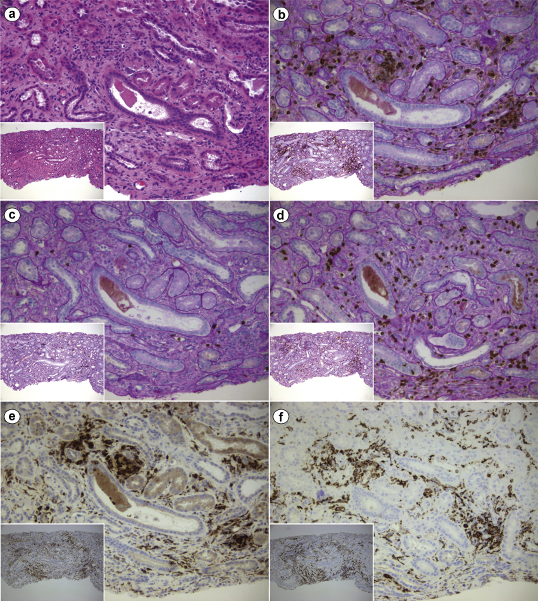
Immunohistochemical characterization of the interstitial inflammatory infiltrate on renal biopsy of a critically ill patient with coronavirus disease 2019 (COVID-19) and acute kidney injury. (a) Mild diffuse interstitial inflammatory infiltrate in the setting of interstitial edema and acute tubular injury with tubuloepithelial flattening and simplification (hematoxylin and eosin, original magnification ×200 and ×100 [inset]). Immunohistochemistry studies showed (b) diffuse leukocyte common antigen (LCA) (CD45)–positive infiltrate (LCA + periodic acid–Schiff [PAS], original magnification ×200 and ×100 [inset]), with (c) a scarce B-cell component (CD20 + PAS, original magnification ×200 and ×100 [inset]), but relevant presence of (d) T cells (CD3 + PAS, original magnification ×200 and ×100 [inset]) and CD68–CD163–positive macrophages (e [CD68] and f [CD163], original magnification ×200 and ×100 [insets]). LCA, as the name implies, is a pan-leukocyte marker; CD3 and C20 are T cell– and B cell–specific, respectively; CD68 and CD163 are both macrophage markers. Overall, the inflammatory infiltrate was mixed and nonspecific, as commonly observed in a variety of conditions, including the typical inflammation associated with tubular atrophy/interstitial fibrosis. CD, cluster of differentiation.
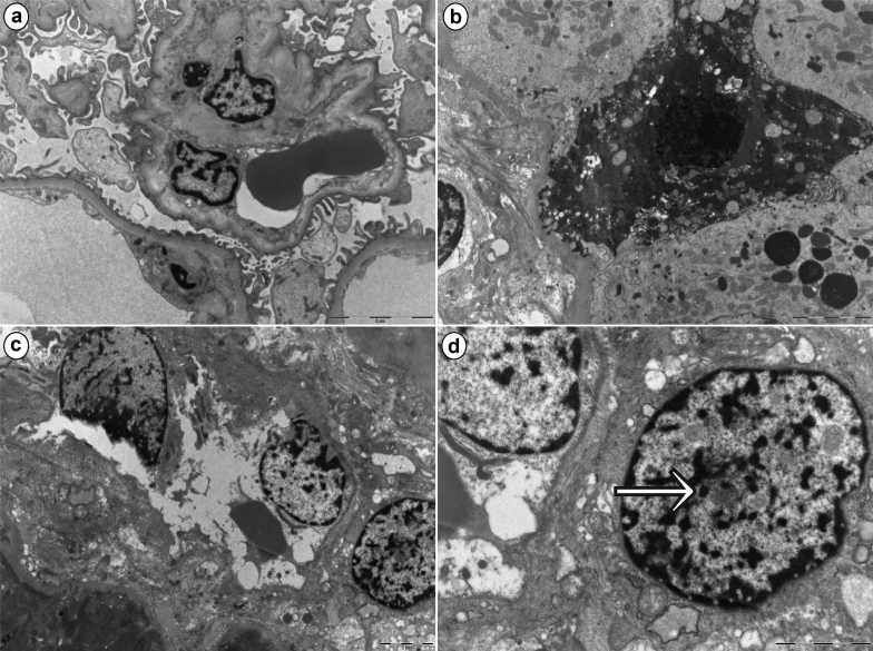
Ultrastructural findings on renal biopsy of a critically ill patient with coronavirus disease 2019 (COVID-19) and acute kidney injury. Low magnification of a glomerulus showing overall well-preserved architecture, with patent capillaries and irregular flattening of podocyte foot processes. (a) Effacement of the latter was minimal and confined to a few areas (bar = 5 μm). (b) A degenerating epithelial cell with a pyknotic nucleus is consistent with acute tubular injury (bar = 5 μm). (c) A blood vessel, likely a peritubular capillary, shows degenerating endothelial cells (bar = 2 μm). Higher magnification of the same image as in (c), highlighting (d) the presence of a “nuclear body” (arrow) in a fibroblast adjacent to the peritubular capillary (bar = 2 μm). Nuclear bodies are thought to be the expression of “cellular hyperactivity” due to several causes, including physiological and disease states.
References
Publication types
LinkOut - more resources
Full Text Sources
Miscellaneous
