. 2020 Aug 3;130(8):4331-4347.
doi: 10.1172/JCI135552.

Selective role of Nck1 in atherogenic inflammation and plaque formation

Affiliations

Selective role of Nck1 in atherogenic inflammation and plaque formation

Mabruka Alfaidi et al. J Clin Invest. .

Abstract

Although the Canakinumab Anti-Inflammatory Thrombosis Outcomes Study (CANTOS) established the role of treating inflammation in atherosclerosis, our understanding of endothelial activation at atherosclerosis-prone sites remains limited. Disturbed flow at atheroprone regions primes plaque inflammation by enhancing endothelial NF-κB signaling. Herein, we demonstrate a role for the Nck adaptor proteins in disturbed flow-induced endothelial activation. Although highly similar, only Nck1 deletion, but not Nck2 deletion, limited flow-induced NF-κB activation and proinflammatory gene expression. Nck1-knockout mice showed reduced endothelial activation and inflammation in both models, disturbed flow- and high fat diet-induced atherosclerosis, whereas Nck2 deletion did not. Bone marrow chimeras confirmed that vascular Nck1, but not hematopoietic Nck1, mediated this effect. Domain-swap experiments and point mutations identified the Nck1 SH2 domain and the first SH3 domain as critical for flow-induced endothelial activation. We further characterized Nck1's proinflammatory role by identifying interleukin 1 type I receptor kinase-1 (IRAK-1) as a Nck1-selective binding partner, demonstrating that IRAK-1 activation by disturbed flow required Nck1 in vitro and in vivo, showing endothelial Nck1 and IRAK-1 staining in early human atherosclerosis, and demonstrating that disturbed flow-induced endothelial activation required IRAK-1. Taken together, our data reveal a hitherto unknown link between Nck1 and IRAK-1 in atherogenic inflammation.

Keywords: Atherosclerosis; Cell Biology; Signal transduction; Vascular Biology; endothelial cells.


Conflict of interest statement

Conflict of interest: The authors have declared that no conflict of interest exists.

Figures

Figure 1
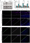
Figure 1. Nck1/2 deletion ameliorates shear stress–induced NF-κB activation.
(A) Human aortic endothelial cells (HAECs) were transfected with siRNA specific for Nck1/2, and transfection efficiency was assessed using Western blotting. (B and C) HAECs were subjected to acute shear stress for the indicated times, and NF-κB activation was assessed by detection of p65 serine 536 phosphorylation using Western blotting. Densitometric analysis was performed using ImageJ. (D and E) p65 nuclear translocation was measured after 45 minutes of shear stress in Nck1/2 siRNA–treated and mock control cells. Images were analyzed using NIS Elements software. Scale bar: 50 μm. Data are presented as the mean ± SEM, n = 4. *P < 0.05, **P < 0.01, ****P < 0.0001 by 2-way ANOVA followed by Bonferroni’s post hoc test. HPF, high-power field. (F and G) HAECs were transfected with Nck1/2 siRNA, and oscillatory shear stress–induced (OSS-induced; ±5 dynes/cm2 with 1 dyne/cm2 forward flow) proinflammatory gene expression (VCAM-1, ICAM-1) and proinflammatory signaling (p-NF-κB Ser536) were assessed by Western blotting. n = 4 in G. *P < 0.05 by Kruskal-Wallis test. (H) HAECs were treated as in F, and mRNA expression was assessed by qRT-PCR (n = 4). ***P < 0.001, ****P < 0.0001 by 2-way ANOVA followed by Bonferroni’s post hoc test.
Figure 2
Figure 2. Nck1, but not Nck2, deletion ameliorates shear stress–induced activation.
(A) Transfection efficiency of selective Nck1 and Nck2 knockdown in HAECs using siRNA. (BE) HAECs lacking either Nck1 or Nck2 were subjected to acute shear stress for the indicated times, and NF-κB activation was assessed by measuring (B and C) NF-κB phosphorylation and (D and E) nuclear translocation. Scale bar: 50 μm. Data are the mean ± SEM, n = 4. (F and G) HAECs were transfected with either Nck1 or Nck2 siRNA, and oscillatory shear stress–induced (OSS-induced, 18 hours) proinflammatory gene expression (ICAM-1/VCAM-1) and signaling (p-NF-κB Ser536) were assessed by Western blotting. (H) Cells transfected as in F and mRNA expression of Klf2, VCAM-1, and ICAM-1 were measured using qRT-PCR. Data are from n = 4 and presented as the mean ± SEM. *P < 0.05, **P < 0.01, ***P < 0.001, ****P < 0.0001 by 2-way ANOVA followed by Bonferroni’s post hoc test.
Figure 3
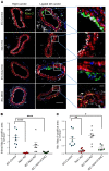
Figure 3. Ablation of Nck1, but not Nck2, blunts partial carotid ligation–induced inflammation.
(A) Schematic of the study in which 4 groups of mice were subjected to the ligation surgery at the indicated time. (B) Endothelial mRNA analysis from iEC-Control, Nck1-KO, iEC-Nck2-KO, and iEC-Nck1/2-DKO mice. mRNA from the ligated left carotid (LC) was normalized to the unligated right carotid (RC) and to the housekeeping gene β-microglobulin. **P < 0.01; ***P < 0.001 by 2-way ANOVA followed by Bonferroni’s post hoc test. (CF) ICAM-1 (red) and VCAM-1 (white) in the ligated left carotid compared with the unligated right carotid arteries among experimental groups. Endothelial cells were stained for von Willebrand factor (vWF in the dashed boxes) and the nuclei counterstained with DAPI. Scale bars: 200 μm (left 2 columns in C and E) and 50 μm (right columns). Images analyzed using NIS Elements software, from n = 7–10 mice/group. Data are the mean ± SEM. **P < 0.01; ***P < 0.001; ****P < 0.0001 by 1-way ANOVA followed by Tukey’s post hoc test.
Figure 4
Figure 4. Nck1 deletion reduces macrophage infiltration in the partial carotid ligation model of disturbed flow.
(A) Macrophages (Mac-2+, green) in the ligated and the unligated carotid arteries among experimental groups. Smooth muscle cells (α-smooth muscle actin+ [SMA+], red), and endothelium (vWF+, white). Scale bars: 200 μm (left 2 columns) and 50 μm (right column). (B and C) Graphical quantification of intimal and adventitial Mac-2–positive cells. Images analyzed using NIS Elements software, from n = 7–10 mice/group. Data are the mean ± SEM. *P < 0.05; **P < 0.01; ****P < 0.0001 by 1-way ANOVA followed by Tukey’s post hoc test.
Figure 5
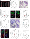
Figure 5. Global deletion of Nck1, but not Nck2, blunts high-fat diet–induced atherosclerosis.
iEC-Control, Nck1-KO, iEC-Nck2-KO, and iEC-Nck1/2-DKO mice were fed high-fat diet (HFD) for 12 weeks. (A) Representative en face morphometric images of total aortic lesion area and (B) calculated whole aortic atherosclerosis (percentage of the total surface area). Scale bar: 1 mm. (C) Representative images of hematoxylin and eosin–stained (H&E-stained) atherosclerotic carotid and (D) quantification of carotid atherosclerotic area among experimental groups. (EG) Analysis of carotid plaque cellular content following staining for macrophages (Mac-2+, green), and smooth muscle cells (α-smooth muscle actin+ [SMA+], red). (H) H&E staining of brachiocephalic arteries (BCAs) after HFD feeding in iEC-Control, Nck1-KO, iEC-Nck2-KO, and iEC-DKO mice. (I) Quantification of atherosclerotic burden in BCAs among experimental groups. (J) Plaque composition assessed by staining for macrophages (Mac-2+) and smooth muscle cells (SMA+) in brachiocephalic lesions. (K) Quantification of Mac-2+ and (L) SMA+ areas in BCAs. Data are the mean ± SEM, n = 6–10/group. *P < 0.05; **P < 0.01; ***P < 0.001; ****P < 0.0001 by 1-way ANOVA followed by Tukey’s post hoc test. Scale bars: 100 μm (C, E, H, and J).
Figure 6
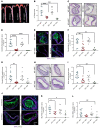
Figure 6. Nck1 deletion from vascular wall, but not hematopoietic cells, has atheroprotective effect.
Nck1-WT (WT) and Nck1-KO mice were irradiated and received bone marrows from either Nck1-WT or Nck1-KO mice. Two weeks after irradiation, the 4 groups of mice were fed HFD for 12 weeks. (A) Representative en face morphometric images of total aortic lesion area and (B) calculated whole aortic atherosclerosis (percentage of the total surface area). Scale bar: 1 mm. (C) Representative images of H&E-stained atherosclerotic carotid artery and (D) quantification of carotid atherosclerotic area among experimental groups. (EG) Analysis of plaque cellular content following staining for macrophages (Mac-2+, green), and smooth muscle cells (α-smooth muscle actin+ [SMA+], purple). (H) H&E staining of brachiocephalic arteries (BCAs) after HFD feeding in WT→WT, WT→KO, KO→WT, and Nck1-KO→Nck1-KO mice. (I) Quantification of atherosclerotic burden in BCAs among experimental groups. (J) Plaque composition assessed by staining for macrophages (Mac-2+) and smooth muscle cells (SMA+) in brachiocephalic lesions. (K) Quantification of Mac-2+ and (L) SMA+ areas in BCAs. Data are the mean ± SEM, n = 4–11/group. *P < 0.05; **P < 0.01; ***P < 0.001; ****P < 0.0001 by 1-way ANOVA and Tukey’s post hoc test. NS, not significant. Scale bars: 100 μm (C, E, H, and J).
Figure 7
Figure 7. Nck1 regulates shear stress–induced inflammation via its SH2 domain and the first of its 3 SH3 domains.
(A) Schematic showing the domain structure of Nck1 and Nck2 and the 2 chimeras consisting of Nck1 SH2/Nck2 SH3.1–SH3.3 and Nck2 SH2/Nck1 SH3.1–SH3.3. (B) Western blot analysis showing comparable transduction efficiency of the Nck1/2 chimeras after transducing the constructs into Nck1/2-DKO cells. n = 3. (C and D) Nck1/2-DKO HAECs were transduced with the constructs in A, and OSS-induced proinflammatory signaling (p-NF-κB Ser536) and gene expression (VCAM-1) were assessed. (E) Schematic of Nck1 SH3 domain point mutations, and (F) Western blot analysis showing the efficiency of reexpression of different Nck1 mutants in Nck1/2-DKO HAECs. (GI) Nck1/2-DKO HAECs were transiently transfected with Nck1 or Nck1 variants described in E, and OSS-induced proinflammatory signaling and gene expression assessed as indicated above. Data are from n = 4–6 and presented as the mean ± SEM. *P < 0.05; **P < 0.01; ***P < 0.001; ****P < 0.0001 by 2-way ANOVA followed by Bonferroni’s post hoc test. NS, not significant.
Figure 8
Figure 8. Nck1 interacts with IRAK1, and IRAK1 activation requires Nck1 in response to shear stress.
(A) Immunoblotting showing Nck1 and IRAK-1 coimmunoprecipitation (Co-IP), confirming Nck1 and IRAK-1 interaction and that it is shear stress dependent. (B and C) Graphical representation of the levels of p-IRAK-1 in Nck-1 Co-IP and Nck1 levels in IRAK-1 Co-IP showing statistical significance after shear stress. Data are from n = 4 and are presented as the mean ± SEM. *P < 0.05, **P < 0.01 by 1-way ANOVA and Tukey’s post hoc test. (D and E) Human aortic endothelial cells (HAECs) were subjected to laminar shear stress (LSS; 10 dynes/cm2, 18 hours) or oscillatory shear stress (OSS; ±5 dynes/cm2 with 1 dyne/cm2 forward flow, 18 hours). Cell lysates were assessed using Western blotting for Nck1, p-IRAK-1, and IRAK-1 levels. GAPDH was used a loading control. Representative blots from n = 4. Data were analyzed by 2-way ANOVA followed by Bonferroni’s post hoc test. (F) Schematic representation of the pathway activation. (G) Lentiviral shRNA–transduced HAECs were subjected to shear stress (±5 dynes/cm2 with 1 dyne/cm2 forward flow, 18 hours). Cell lysates were assessed using Western blotting for p-IRAK-1 and IRAK-1 levels. GAPDH was used a loading control. Representative blots from n = 4; data presented as the mean ± SEM. **P < 0.01 by 2-way ANOVA followed by Bonferroni’s post hoc test. (H) Immunostained ligated left carotid arteries from iEC-Control, Nck1-KO, iEC-Nck2-KO, and iEC-Nck1/2-DKO mice. p-IRAK-1 (red), predominately within the endothelium (vWF+, green), is reduced in Nck1-KO mice. Scale bar: 200 μm. (I) Graphical quantification showing the significance of the data from H. Data presented as the mean ± SEM. ***P < 0.001;****P < 0.0001 by 1-way ANOVA and Tukey’s post hoc test.
Figure 9
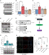
Figure 9. IRAK1 deletion ameliorates shear stress–induced activation.
(A) HAECs were treated with or without IRAK-1 siRNA and subjected to oscillatory shear stress (OSS, 18 hours). Cell lysates were analyzed for VCAM-1, ICAM-1, and p-NF-κB using Western blotting. (B) Quantification of ICAM-1, VCAM-1, and p-NF-κB showing significant reduction in IRAK-1–depleted cells. Densitometric analysis was performed using ImageJ. Data are presented as the mean ± SEM. ***P < 0.001, ****P < 0.0001 by 2-way ANOVA followed by Bonferroni’s post hoc test. (C) Type I human atherosclerotic lesions were stained for p-IRAK-1 and Nck1, and vWF was used as an endothelial marker. (D) Healthy coronary sections were stained for p-IRAK-1, Nck1, and vWF. Dashed marks indicate internal elastic lamina. Scale bars: 100 μm. Images analyzed using NIS Elements software, from n = 5 postmortem biopsy samples.

Comment in

References

    1. Libby P, Ridker PM, Hansson GK, Leducq Transatlantic Network on Atherothrombosis Inflammation in atherosclerosis: from pathophysiology to practice. J Am Coll Cardiol. 2009;54(23):2129–2138. doi: 10.1016/j.jacc.2009.09.009. - DOI - PMC - PubMed
    1. Dai GH, et al. Distinct endothelial phenotypes evoked by arterial waveforms derived from atherosclerosis-susceptible and -resistant regions of human vasculature. Proc Natl Acad Sci U S A. 2004;101(41):14871–14876. doi: 10.1073/pnas.0406073101. - DOI - PMC - PubMed
    1. Davies PF. Flow-mediated endothelial mechanotransduction. Physiol Rev. 1995;75(3):519–560. doi: 10.1152/physrev.1995.75.3.519. - DOI - PMC - PubMed
    1. Ross R. Atherosclerosis--an inflammatory disease. N Engl J Med. 1999;340(2):115–126. doi: 10.1056/NEJM199901143400207. - DOI - PubMed
    1. Brooks AR, Lelkes PI, Rubanyi GM. Gene expression profiling of human aortic endothelial cells exposed to disturbed flow and steady laminar flow. Physiol Genomics. 2002;9(1):27–41. doi: 10.1152/physiolgenomics.00075.2001. - DOI - PubMed

Publication types

MeSH terms