. 2020 May 19;10(1):8227.
doi: 10.1038/s41598-020-64940-2.

Assessment of a pro-healing stent in an animal model of early neoatherosclerosis

Affiliations

Assessment of a pro-healing stent in an animal model of early neoatherosclerosis

Philipp Nicol et al. Sci Rep. .

Abstract

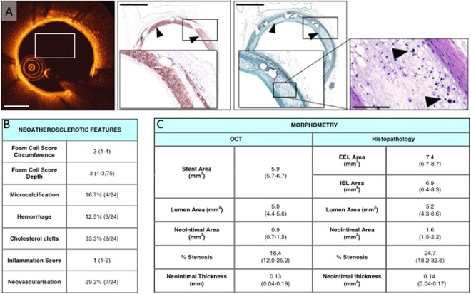
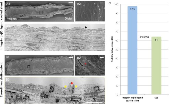
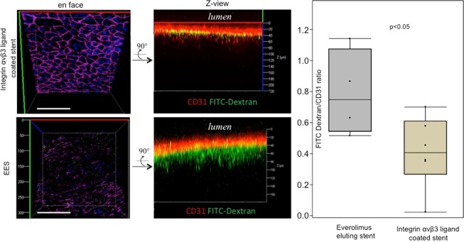
Background: Neoatherosclerosis represents an accelerated manifestation of atherosclerosis in nascent neointima after stenting, associated with adverse events. We investigated whether improved reendothelialization using RGD-coated stents results in diminished vascular permeability and reduced foam cell formation compared to standard DES in atherosclerotic rabbits.

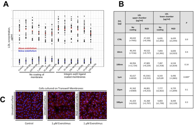
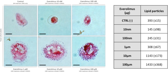
Methods and results: Neointimal foam cell formation was induced in rabbits (n = 7). Enhanced endothelial integrity in RGD-coated stents resulted in decreased vascular permeability relative to DES, which was further confirmed by SEM and TEM. Cell culture experiments examined the effect of everolimus on endothelial integrity. Increasing concentrations of everolimus resulted in a dose-dependent decrease of endothelial cell junctions and foam cell transformation of monocytes, confirming the relevance of endothelial integrity in preventing permeability of LDL.

Conclusion: Incomplete endothelial integrity was confirmed as a key factor of neointimal foam cell formation following stent implantation. Pro-healing stent coatings may facilitate reendothelialization and reduce the risk of neoatherosclerosis.


Conflict of interest statement

Dr. Joner reports grants from ESC Grant for Medical Research Innovation, during the conduct of the study; personal fees from Consulting for Biotronik, personal fees from Speakers’ fee from Biotronik, personal fees from Consulting for Orbus Neich, personal fees from Speakers’ fee from Orbus Neich, personal fees from Speakers’ fee from Boston Scientific, personal fees from Speakers’ fee from Medtronic, personal fees from Speakers’ fee from Astra Zeneca, outside the submitted work. All other authors have no conflicts of interest to declare.

Figures

Figure 1
Figure 1
(A) ProKinetic Energy BMS in a rabbit iliac artery 12 weeks after implantation, assessed by histopathology and OCT. OCT shows surface with almost circumferential high backscattering intensity and attenuation. Corresponding histological cross section (Movat Pentachrome staining) shows circumferential foamy macrophage accumulation in a moderately thickened neointimal tissue (arrowheads indicate foamy macrophages; scale bar = 1000 µm). High-magnified image of Hematoxylin Eosin staining shows microcalcifications between foamy macrophages (scale bar = 100 µm). (B) Neointimal characteristics from study 1 and (C) morphometric analysis derived from OCT and histopathology (n = 5 rabbits, 24 quadrants scored in total).
Figure 2
Figure 2
Comparison of integrin αvβ3 ligand coated stent (A1–A3) and EES (B1–B3) 12 weeks after implantation in a hypercholesterolemic rabbit model with quantification of endothelial coverage (C). Scanning electron microscopy (SEM) of an integrin αvβ3 ligand coated stent half (A1) and an Everolimus eluting stent (EES) half (B1) shows improved strut-coverage as compared to EES. High-magnification SEM images (A2 and B2) confirm a continuous monolayer of endothelial cells above integrin αvβ3 ligand coated stent struts whereas EES-struts seem to be covered by loosely arranged endothelial cells in the presence of scattered inflammatory cells and platelets (red asterisks = stent strut). Transmission electron microscopy (TEM) demonstrates a continuous endothelial monolayer with abundant intercellular junctions (arrowheads) in an integrin αvβ3 ligand coated stent (A3) while impaired endothelial monolayer integrity is observed in EES (B3, yellow arrowheads mark endothelial cells in the absence of intercellular junction, red arrowhead indicates incidental finding of a transmigrating monocyte). Scale bar: A1/B1 = 1 mm. A2/A3 = 25 µm. A3/B3 = 100 µm.
Figure 3
Figure 3
Left: Selective confocal microscopy images of an integrin αvβ3 ligand coated stent (top) and EES (bottom) 12 weeks after implantation in a hypercholesterolemic rabbit model. En face images (left) show strong CD31 staining of endothelial cells (cell shape, red channel, pink pseudocolor) in the integrin αvβ3 ligand coated stent and decreased CD31 staining in EES. FITC-dextran accumulation (green channel) between endothelial cells (red channel) is increased in EES as compared to integrin αvβ3 ligand coated stents (p < 0.05). n = 5 each and expressed as means with standard deviation calculated by ANOVA. Scale bar = 1 mm.
Figure 4
Figure 4
(A,B) AcLDL-concentrations in an in vitro permeability assay (transwell model) above and below endothelium which was cultured on ± integrin αvβ3 ligand coated semipermeable membranes and treated with everolimus in different concentrations (see B) for 24 h. Everolimus treatment causes a dose-dependent decrease of AcLDL in the upper compartment of the semipermeable membrane and an increase of AcLDL in the lower compartment (mean LDL concentration above endothelium marked in red and below endothelium in blue; n = 15). (C) Endothelial cells cultured on transwell membranes exposed to different concentrations of everolimus. The control group (uncoated surface) shows a confluent monolayer with intense VE-Cadherin staining (left). Incubation with everolimus at 1 µM for 24 h resulted in incomplete endothelial integrity on uncoated surfaces (centre image). Endothelial cells cultured on integrin αvβ3 ligand coated surfaces (right image) show preserved VE-Cadherin expression and less intercellular gaps (For results on quantification, please see Supplemental Fig. 3). Scale bar = 100 µm.
Figure 5
Figure 5
Brightfield images of foamy monocytes in the presence of AcLDL (24 h incubation on tissue culture plastic, n = 3) and results of automated quantification of lipid particles. Monocytes were co-cultured with endothelial cells after exposure to everolimus. Increasing concentrations of everolimus and fixed concentration of AcLDL result in dose-dependent transformation of monocytes into foam cells. (Foam cells stained with Oil-red-O, greater lipid accumulation in monocytes co-cultured with endothelial cells under high concentrations of everolimus). Scale bar = 20 µm.

References

    1. Serruys PW, et al. A comparison of balloon-expandable-stent implantation with balloon angioplasty in patients with coronary artery disease. Benestent Study Group. N. Engl. J. Med. 1994;331:842–845. doi: 10.1056/NEJM199408253310801. - DOI - PubMed
    1. Fischman D, et al. A randomized comparison of coronary-stent placement and balloon angioplasty in the treatment of coronary artery disease. Stent Restenosis Study Investigators. N. Engl. J. Med. 1994;331:842–845. doi: 10.1056/NEJM199408253310802. - DOI - PubMed
    1. Foley DP, et al. Differences in restenosis propensity of devices for transluminal coronary interventionA quantitative angiographic comparison of balloon angioplasty, directional atherectomy, stent implantation and excimer laser angioplasty. Eur. Heart J. 1995;16:1331–1346. doi: 10.1093/oxfordjournals.eurheartj.a060740. - DOI - PubMed
    1. Nakazawa G, et al. The pathology of neoatherosclerosis in human coronary implants: Bare-metal and drug-eluting stents. J. Am. Coll. Cardiol. 2011;57:1314–1322. doi: 10.1016/j.jacc.2011.01.011. - DOI - PMC - PubMed
    1. Finn AV, et al. Pathological correlates of late drug-eluting stent thrombosis: Strut coverage as a marker of endothelialization. Circulation. 2007;115:2435–2441. doi: 10.1161/CIRCULATIONAHA.107.693739. - DOI - PubMed

Publication types