. 2020 May 19;10(1):8250.
doi: 10.1038/s41598-020-64931-3.

Induction of posterior vitreous detachment (PVD) by non-enzymatic reagents targeting vitreous collagen liquefaction as well as vitreoretinal adhesion

Affiliations

Induction of posterior vitreous detachment (PVD) by non-enzymatic reagents targeting vitreous collagen liquefaction as well as vitreoretinal adhesion

Mithun Santra et al. Sci Rep. .

Erratum in

Abstract

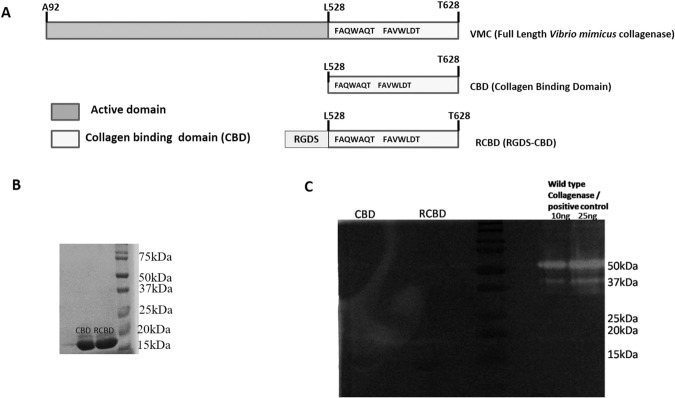
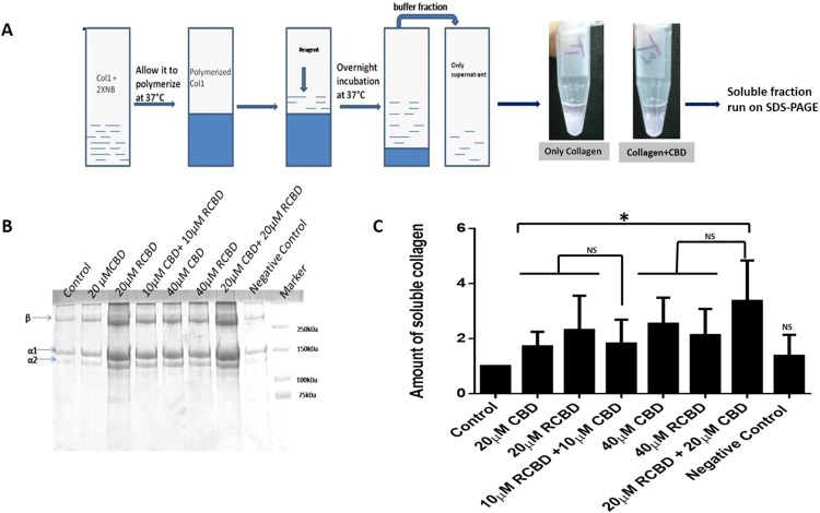
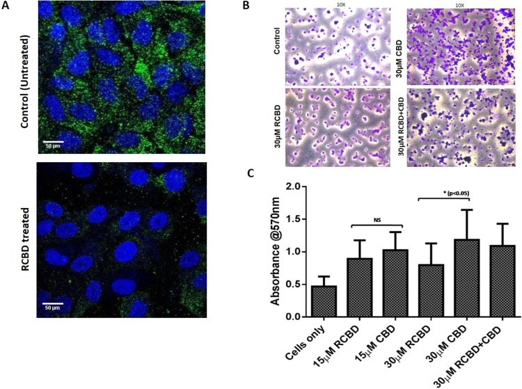
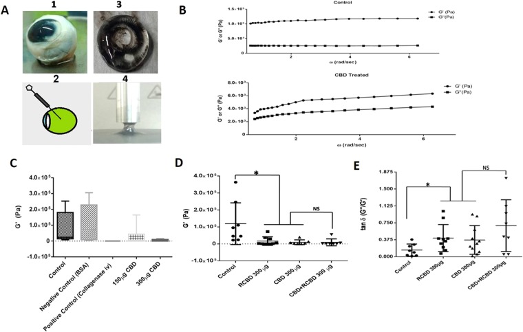
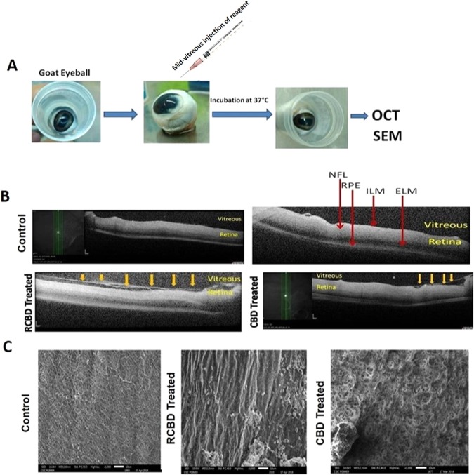
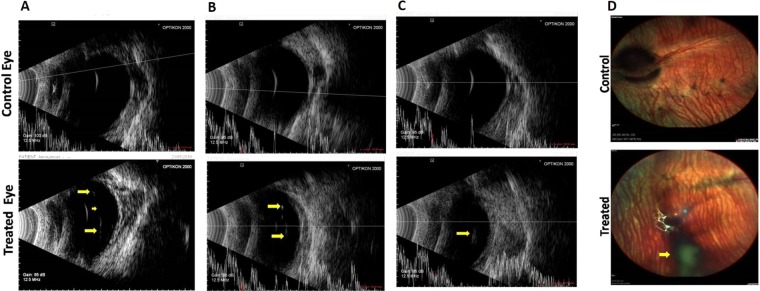
Induction of posterior vitreous detachment (PVD) by pharmacologic vitreolysis has been largely attempted through the use of enzymatic reagents. Ocriplasmin has been the only FDA-approved clinical reagent so far. Several adverse effects of ocriplasmin have emerged, however, and the search for alternative PVD-inducing reagents continues. Since i) collagen forms an important structural component of the vitreous, and ii) strong vitreo-retinal adhesions exist between the cortical vitreous and the internal limiting membrane (ILM) of the retina, an effective PVD-inducing reagent would require both, vitreous liquefaction, and concurrent dehiscence of vitreoretinal adhesion, without being toxic to retinal cells. We designed a combination of two reagents to achieve these two objectives; a triple helix-destabilizing collagen binding domain (CBD), and a fusion of RGD (integrin-binding) tripeptide with CBD (RCBD) to facilitate separation of posterior cortical vitreous from retinal surface. Based on in vitro, ex-vivo, and in vivo experiments, we show that a combination of CBD and RCBD displays potential for safe pharmacologic vitreolysis. Our findings assume significance in light of the fact that synthetic RGD-containing peptides have already been used for inhibition of tumor cell invasion. Proteins such as variants of collagen binding domains could have extended therapeutic uses in the future.


Conflict of interest statement

The authors declare no competing interests.

Figures

Figure 1
Figure 1
(Panel A) Schematic representation of vibrio mimicus collagenase (VMC)-derived constructs; (Panel B) SDS-PAGE profile of the proteins, CBD and RCBD; (Panel C) Zymography showing no gelatinolytic activity of CBD or RCBD. Full-length gel is presented in Supplementary Fig. S2.
Figure 2
Figure 2
Secondary structure determination by circular dichroism (CD). CD spectra of CBD and RCBD (Panel A) and VMC (Panel B).
Figure 3
Figure 3
Structural perturbation of collagen upon binding of CBD and RCBD. Mean residue ellipticity (MRE) vs wavelength plots for collagen type I incubated with CBD (Panels A-B) and RCBD (Panels D-E) at two different concentrations (2.5 µM and 5 uM) at 30 °C, for 0 h, 1 h and 3 h; similar plots for col I incubated with varying concentrations of CBD (Panel C) and RCBD (Panel F) at 30 °C after 3 h of incubation with the respective reagents.
Figure 4
Figure 4
Collagen solubilization assay. (Panel A) Experimental outline of the protocol; collagen type 1 was polymerized in the presence of neutralizing buffer, followed by treatment with the test reagents, and incubated overnight at room temperature; the supernatant from each tube was run on SDS-PAGE to assess the released monomeric soluble collagen. A representative image showing decreased opacity of the polymerized collagen in the presence of CBD as compared to the control tube (minus CBD); (Panel B) Representative SDS-PAGE gel of the assay; (Panel C) Upon treatment with CBD/RCBD, polymerized collagen releases the monomeric form of (soluble) collagen, seen to be increased in samples treated with reagents (indicated by increased band intensity of α1 and α2 chains of collagen), as compared to untreated sample of collagen (control). Negative control: 20 μM CK (cysteine knot domain in the C-terminal region of CTGF); Data represents the mean (± standard deviation, SD) of five independent experiments (*p < 0.05, significant; NS, Not Significant).
Figure 5
Figure 5
RCBD binds to cell surface integrins and causes decreased adherence of ARPE-19 cells to collagen as compared to CBD: (Panel A) Immunofluorescence staining of β1 integrin in ARPE-19 cells. Cells were seeded on coverslips in presence of vitreous derived from patients undergoing surgery for retinal detachment, in the absence and presence of RCBD; immunofluorescence staining was done using mouse anti-β1 integrin antibody and goat anti-mouse-FITC secondary antibody. β1 integrin-associated fluorescence is shown in green and nuclei (DAPI) in blue; scale bar: 50 µm (Panel B) Adherence of ARPE-19 cells (untreated or pre-treated with CBD and/or RCBD) on collagen coated surface; adhered cells were imaged; scale bar: 400 µm, 10X magnification; and (Panel C) Bar diagrams represent the corresponding quantitative data.
Figure 6
Figure 6
Rheological studies of goat eye vitreous treated with CBD/RCBD vitreous: (Panel A) Experimental set-up; (Panel B) Measurements of storage modulus (G’) and loss modulus (G”) were done for the control vitreous sample (treated with PBS) and vitreous treated with reagents. Frequency sweeps were recorded at fixed shear strain amplitude (γ○ =3%); linear viscoelastic region (i.e., modulus independent of frequency) is taken within the range of π/5 to 2π rad/s of oscillatory frequency sweep. Error bars indicate standard deviation; (Panel C) Upon treatment of vitreous with CBD, G’ decreased in a dose-dependent manner whereas injection with PBS (control) or BSA (negative control) did not show any change in G’; (D) Both, CBD as well as RCBD-treated vitreous resulted in a significant decrease in storage modulus (P < 0.05). The combination of CBD and RCBD showed a similar effect as seen with individual reagents; (E) The loss tangent (G”/G’) increased significantly in the presence of either reagent used individually, as well as in combination, as compared to control (p < 0.05). (**No of goat eyeballs per treatment group, n = 3).
Figure 7
Figure 7
Ex-vivo OCT and SEM studies in goat eyeballs: (Panel A) Schematic representation of the experimental outline; (Panel B) Representative OCT images showing detachment of posterior hyaloid membrane of vitreous from the ILM of the retina upon treatment with 250 µg CBD or 350 µg RCBD (shown by arrows); no posterior vitreous detachment (PVD) was seen in the corresponding contralateral eyes; (Panel C) SEM micrographs showing dense vitreous fibrils on the retinal surface of control eye, which were cleared/disorganized upon treatment with reagents (**scale bar: 10 µm); (ELM: External Limiting Membrane, ILM: Inner Lining Membrane, NFL: Nerve Fiber Layer, RPE: Retinal Pigment Epithelial cells).
Figure 8
Figure 8
B-scan ultrasonography in three representative eyes following treatment with RCBD + CBD: Representative images of eyes treated with CBD and RCBD showing complete PVD (Panels A and B, marked by arrows), and partial PVD (Panel C), along with the corresponding contralateral eyes (controls). (Panel D) Fundus of representative rabbit eye with complete PVD, following the intravitreal injection of combination of CBD and RCBD; the posterior hyaloid membrane appears as a shadow in the fundus image which reflects PVD (marked with arrow).
Figure 9
Figure 9
Scanning electron photomicrographs of the inner retinal surface of 3 representative animals treated with CBD and RCBD. In test eyes with complete PVD (Panels A and B), the retinal surface was devoid of any collagen fibers; in eyes with partial PVD (Panel C), there was less vitreous cortex left on the retinal surface, compared to the corresponding control (contralateral) eyes (PBS) with no PVD, where dense vitreous cortical fibers were visible (** Scale Bar 1 µm or 10000X magnification).

References

    1. Lund-Andersen H, Sebag J, Sander B, La Cour M. The vitreous. Advances in Organ Biology. 2005;10:181–194. doi: 10.1016/S1569-2590(05)10007-X. - DOI
    1. Sebag J. Ageing of the vitreous. Eye. 1987;1:254. doi: 10.1038/eye.1987.45. - DOI - PubMed
    1. Bishop PN. Structural macromolecules and supramolecular organisation of the vitreous gel. Prog. Retin. Eye Res. 2000;19:323–44. doi: 10.1016/S1350-9462(99)00016-6. - DOI - PubMed
    1. Le Goff MM, Bishop PN. Adult vitreous structure and postnatal changes. Eye. 2008;22:1214–22. doi: 10.1038/eye.2008.21. - DOI - PubMed
    1. De Smet MD, Elkareem AM, Zwinderman AH. The vitreous, the retinal interface in ocular health and disease. Ophthalmologica. 2013;230:165–78. doi: 10.1159/000353447. - DOI - PubMed

Publication types

MeSH terms