

Comparison of Three Histological Techniques for Fat Emboli Detection in Lung Cetacean's Tissue
- PMID: 32427895
- PMCID: PMC7237497
- DOI: 10.1038/s41598-020-64821-8
Comparison of Three Histological Techniques for Fat Emboli Detection in Lung Cetacean's Tissue
Abstract
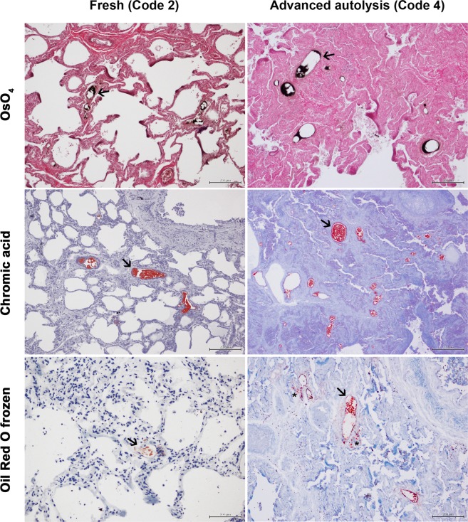
Fat embolism is the mechanical blockage of blood vessels by circulating fat particles. It is frequently related to traumas involving soft tissues and fat-containing bones. Different techniques have been used for decades to demonstrate histologically fat emboli, being the extremely toxic post-fixation with osmium tetroxide one of the most used techniques in the last decades. In the present study, the osmium tetroxide technique was compared qualitatively and quantitatively, for the first time, with chromic acid and Oil Red O frozen techniques for histological fat emboli detection in the lungs of eight sperm whales that died due to ship strikes. This was also the first time that chromic acid technique was tested in cetaceans. Results showed that the three techniques were valuable for the histological detection of fat embolism in cetaceans, even when tissues presented advanced autolysis and had been stored in formaldehyde for years. Although quantitative differences could not be established, the Oil Red O frozen technique showed the lowest quality for fat emboli staining. On the contrary, the chromic acid technique was proven to be a good alternative to osmium tetroxide due to its slightly lower toxicity, its equivalent or even superior capacity of fat emboli detection, and its significantly lower economic cost.
Conflict of interest statement
The authors declare no competing interests.
Figures
References
-
- Levy D. The Fat Embolism Syndrome. Clin. Orthop. Relat. Res. 1990;261:281–286. - PubMed
Publication types
MeSH terms
Grants and funding
LinkOut - more resources
Full Text Sources
Medical
