

Case Reports
doi: 10.36660/abc.20190487.
Epub 2020 May 18.
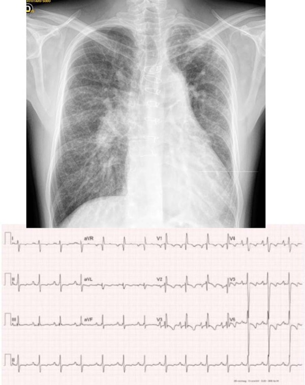
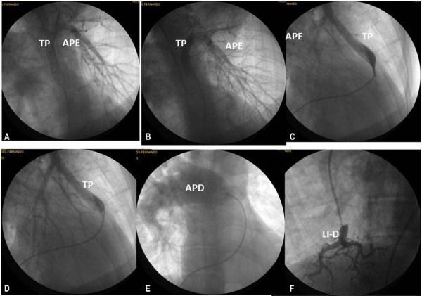
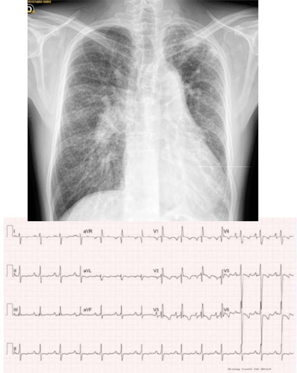
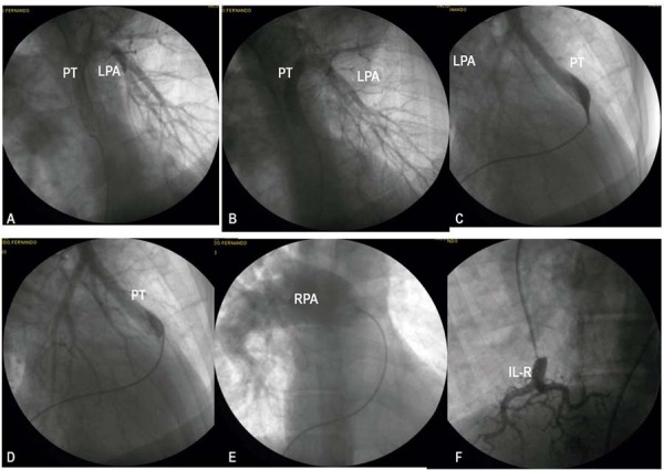
Case 3/2020 - Pulmonary Atresia, Interventricular Communication and Anomalous Origin of the Right Pulmonary Artery from the Ascending Aorta developing after Prior Left Central Shunt, in a Symptomatic 40-year-old Adult
[Article in
English,
Portuguese]
Affiliations
- PMID: 32428103
- PMCID: PMC8149107
- DOI: 10.36660/abc.20190487
Item in Clipboard
Case Reports
Case 3/2020 - Pulmonary Atresia, Interventricular Communication and Anomalous Origin of the Right Pulmonary Artery from the Ascending Aorta developing after Prior Left Central Shunt, in a Symptomatic 40-year-old Adult
[Article in
English,
Portuguese]
Arq Bras Cardiol.
2020 Apr.
No abstract available
Figures
Similar articles
-
A rare case of anomalous origin of the left pulmonary artery from the ascending aorta with ventricular septal defect and pulmonary atresia.Kardiol Pol. 2023;81(10):1022-1023. doi: 10.33963/KP.a2023.0172. Epub 2023 Aug 4. Kardiol Pol. 2023. PMID: 37537919 No abstract available.
-
A rare case of tetralogy of Fallot-pulmonary atresia with absent right pulmonary artery and anomalous origin of left pulmonary artery from ascending aorta.J Card Surg. 2022 Jun;37(6):1718-1719. doi: 10.1111/jocs.16455. Epub 2022 Mar 26. J Card Surg. 2022. PMID: 35338714
-
Pulmonary atresia and ventricular septal defect with collaterals to right lung associated with anomalous left pulmonary artery from the ascending aorta.Pediatr Radiol. 2010 Dec;40 Suppl 1:S72-6. doi: 10.1007/s00247-010-1832-2. Epub 2010 Sep 24. Pediatr Radiol. 2010. PMID: 20865412
-
Anomalous right pulmonary artery from aorta, surgical approach case report and literature review.J Card Surg. 2021 Aug;36(8):2890-2900. doi: 10.1111/jocs.15618. Epub 2021 May 28. J Card Surg. 2021. PMID: 34047395 Free PMC article. Review.
-
Pulmonary Atresia With an Intact Ventricular Septum: Preoperative Physiology, Imaging, and Management.Semin Cardiothorac Vasc Anesth. 2018 Sep;22(3):245-255. doi: 10.1177/1089253218756757. Epub 2018 Feb 7. Semin Cardiothorac Vasc Anesth. 2018. PMID: 29411679 Review.
References
-
- 1. Pepeta L, Takawira FF, Cilliers AM, Adams PE, Ntsinjana NH, Mitchell BL. Anomalous origin of the left pulmonary artery from the ascending aorta in two children with pulmonary atresia, sub-aortic ventricular septal defect and right-sided major aorto-pulmonary collateral arteries. Cardiovasc J Afr. 2011; 22(5): 268-71. - PubMed
-
- 2. Makhmudov MM, abdumadzhidov KhA, Abdullaev EE, Mirzakhmedov BM. A case of tetralogy of Fallot with atresia of the pulmonary trunk and origin of the left pulmonary artery from the ascending aorta. Grudn Khir. 1987 Nov/Dec;(6):82-3. - PubMed
-
- 3. Metras DR, Kreitmann B, Tatou E, Riberi A, Werment F. Tetralogy of Fallot with pulmonary atresia, coronary artery-pulmonary artery fistula, and origin of left pulmonary artery from descending aorta: total correction in infancy. J Thorac Cardiovasc Surg. 1993;105(1):186-8. - PubMed
Publication types
MeSH terms
LinkOut - more resources
Full Text Sources
Medical
