

Comment
doi: 10.1093/brain/awaa120.
A homozygous GDAP2 loss-of-function variant in a patient with adult-onset cerebellar ataxia
Affiliations
- PMID: 32428220
- PMCID: PMC7296852
- DOI: 10.1093/brain/awaa120
Item in Clipboard
Comment
A homozygous GDAP2 loss-of-function variant in a patient with adult-onset cerebellar ataxia
Brain.
.
No abstract available
Figures
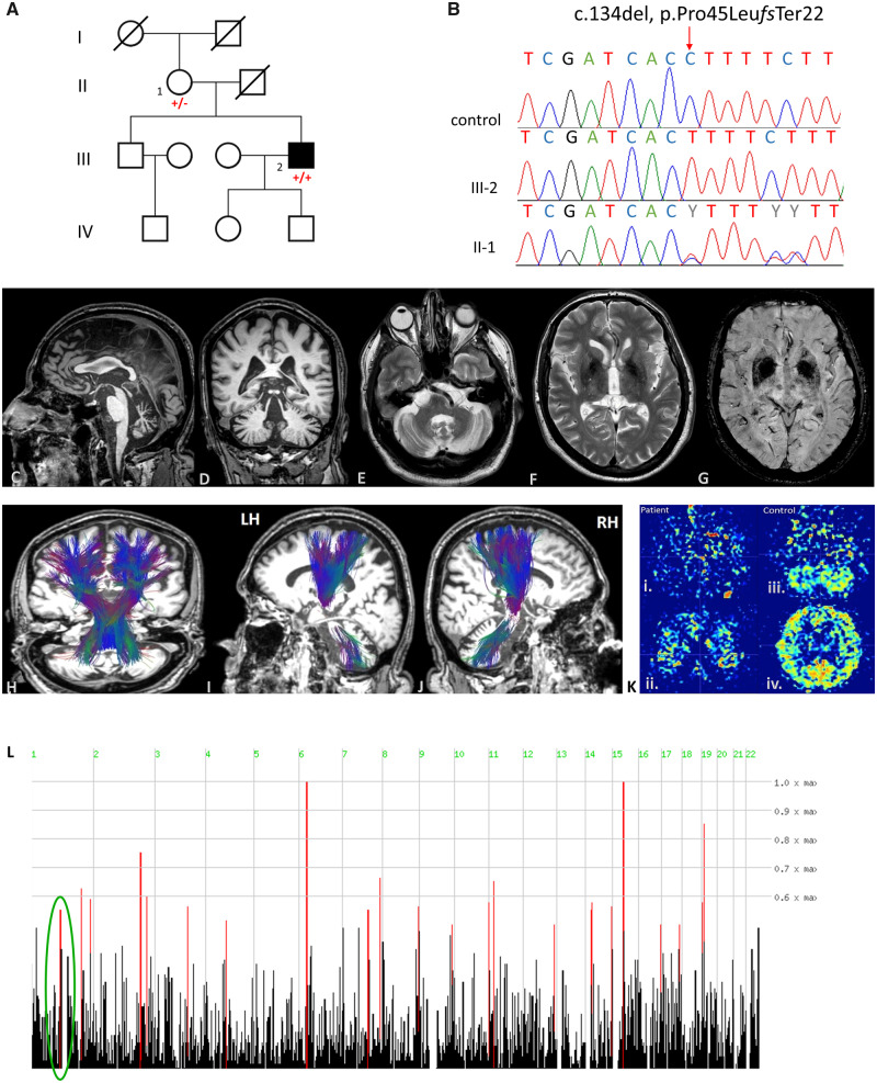
Genetic and neuroimaging findings of this case: (A) Family pedigree. (B) Validation of the variant identified (c.134del, p.Pro45Leufs*22) by Sanger sequencing as homozygous in the patient and heterozygous in the mother compared to a control reference. (C–J) Brain MRI findings of this case. (C) Sagittal T2-weighted sequence showing cerebellar atrophy, midbrain and pons atrophy (hummingbird sign), and thinning of corpus callosum. (D and E) Coronal and axial T1-weighted sequence. Cerebellar atrophy is noted particularly in upper and central cerebellar vermis, however cortical atrophy, and dilated fissures (Global Cortical Atrophy, GCA scale: 2) were also present. (F and G) Axial T2-weighted and SWI (susceptibility weighted imaging) sequences showing lentiform hemosiderin depositions. (H–J) Magnetic resonance tractography using diffusion tensor imaging shows no significant alterations in the reconstructed corticospinal and cerebellar tracts superimposed on anatomical 3D T1-weighted images in axial, coronal and sagittal planes. L = left; R = right; LH = left hemisphere; RH = right hemisphere. [K(i and ii)] Patient’s ASL (arterial spin labelling) representative colour-coded images at cerebellar and lateral ventricles level showing reduced perfusion in cerebellum and in cerebral cortex. [K(iii and iv)] Control ASL representative colour coded images at cerebellar and lateral ventricles level. (L) Homozygosity mapping showing homozygous segments on chromosome 1 where the variant identified resides within a 3.1 Mb homozygous chromosomal segment (ch1: 114 522 285–117 628 869).
Comment in
-
Reply: A homozygous GDAP2 loss-of-function variant in a patient with adult-onset cerebellar ataxia; and Novel GDAP2 pathogenic variants cause autosomal recessive spinocerebellar ataxia-27 (SCAR27) in a Chinese family.Brain. 2020 Jun 1;143(6):e51. doi: 10.1093/brain/awaa122. Brain. 2020. PMID: 32428197 Free PMC article. No abstract available.
Comment on
-
GDAP2 mutations implicate susceptibility to cellular stress in a new form of cerebellar ataxia.Brain. 2018 Sep 1;141(9):2592-2604. doi: 10.1093/brain/awy198. Brain. 2018. PMID: 30084953 Free PMC article.
References
-
- Gonzalez Esquivel D, Ramirez-Ortega D, Pineda B, Castro N, Rios C, Perez de la Cruz V.. Kynurenine pathway metabolites and enzymes involved in redox reactions. Neuropharmacology 2017; 112: 331–45. - PubMed
