Review
. 2020 May 18;30(10):R535-R543.
doi: 10.1016/j.cub.2020.03.047.

Tracing the Evolutionary Origin of Desmosomes

Affiliations
Review

Tracing the Evolutionary Origin of Desmosomes

Kathleen J Green et al. Curr Biol. .

Abstract

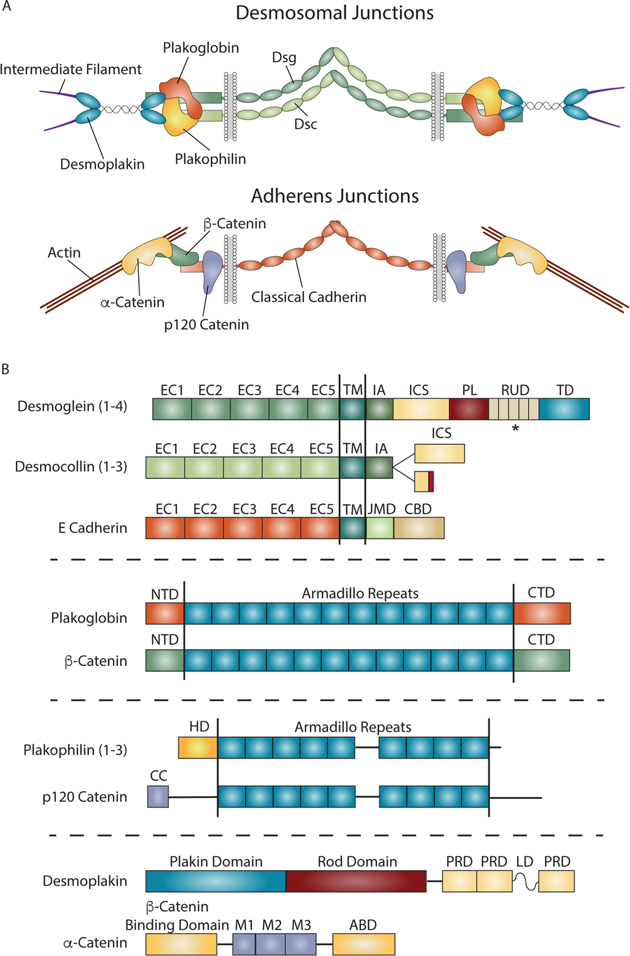
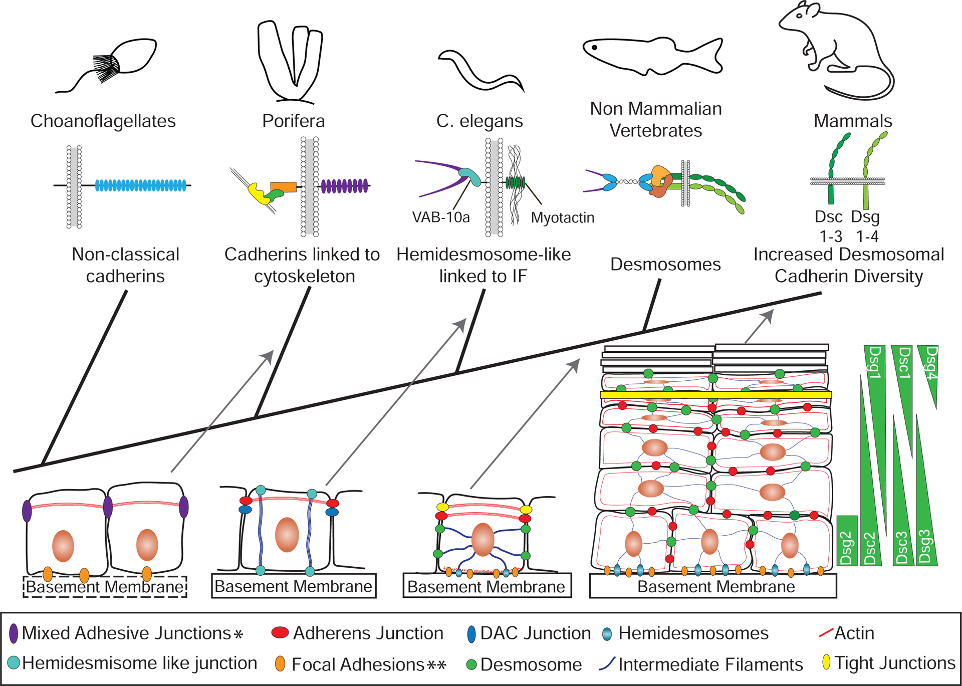
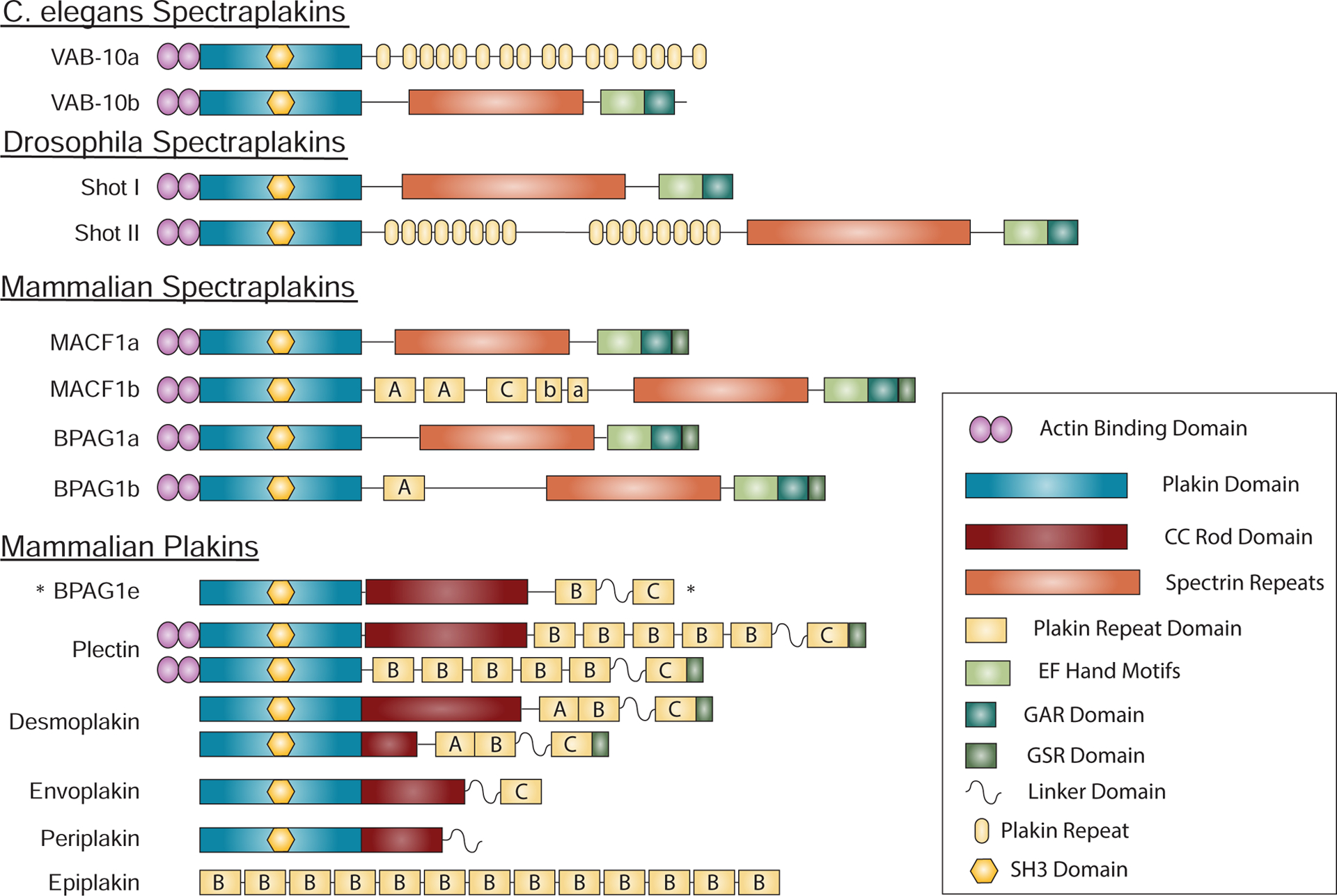
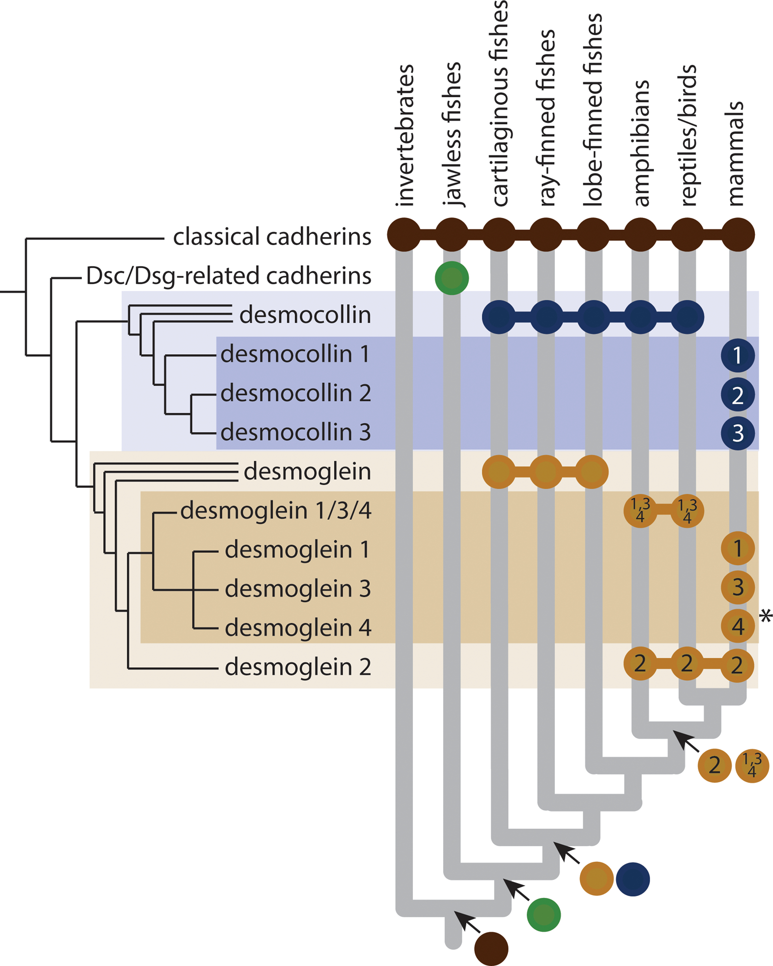
Cadherin-based cell-cell junctions help metazoans form polarized sheets of cells, which are necessary for the development of organs and the compartmentalization of functions. The components of the protein complexes that generate cadherin-based junctions have ancient origins, with conserved elements shared between animals as diverse as sponges and vertebrates. In invertebrates, the formation and function of epithelial sheets depends on classical cadherin-containing adherens junctions, which link actin to the plasma membrane through α-, β- and p120 catenins. Vertebrates also have a new type of cadherin-based intercellular junction called the desmosome, which allowed for the creation of more complex and effective tissue barriers against environmental stress. While desmosomes have a molecular blueprint that is similar to that of adherens junctions, desmosomal cadherins - called desmogleins and desmocollins - link intermediate filaments (IFs) rather than actin to the plasma membrane through protein complexes comprising relatives of β-catenin (plakoglobin) and p120 catenin (plakophilins). In turn, desmosomal catenins interact with members of the IF-binding plakin family to create the desmosome-IF linking complex. In this Minireview, we discuss when and how desmosomal components evolved, and how their ability to anchor the highly elastic and tough IF cytoskeleton endowed vertebrates with robust tissues capable of not only resisting but also properly responding to environmental stress.


Figures

Figure 1.
Figure 1.. Comparison of Desmosome and Adherens Junction Composition and Structure.
A) Both desmosomal and adherens junctions are built from cadherins and associated catenins that facilitate linkage to intermediate filaments (IF) and actin, respectively. In desmosomal junctions, adhesion is mediated by interactions between the ectodomains of desmosomal cadherins, desmogleins (Dsg) and desmocollins (Dsc). The desmosomal cadherin cytoplasmic domains anchor IF to the membrane through protein complexes containing plakoglobin and plakophilins, and the plakin desmoplakin. Adherens junctions mediate intercellular adhesion through homophilic interactions between cadherin ectodomains and link actin to the plasma membrane through α-, β- and p120 catenins that associate with the cadherin cytodomain. B) Domain structures for the major components of the desmosome and adherens junctions. The domain structures of desmoglein and desmocollin are compared to the classical cadherin E-cadherin. All three cadherins contain five extracellular cadherin (EC) domains responsible for adhesion, followed by a single pass transmembrane domain (TM), and a cytoplasmic tail domain that interacts with cytoskeletal linking complexes. The cytoplasmic domains contain an intracellular anchor (IA) and intracellular cadherin-like sequence (ICS) in desmosomal cadherins, and a juxtamembrane domain (JMD) and catenin binding domain (CBD) in classical cadherins. The ICS and CBD domains associate with plakoglobin or β-catenin/plakoglobin, in desmosomal and classical cadherins, respectively. Desmocollins exist as two splice variants, with the Dsc a form containing a full ICS domain, while Dsc b contains a truncated ICS domain and a short sequence of unique amino acids at the C-terminus. Desmogleins also contain an extended tail sequence with an intracellular proline rich linker (PL) domain, repeat unit domain (RUD) with varying number of repeats, and a desmoglein terminal domain (TD). Plakoglobin and β-catenin are armadillo proteins in desmosomes and adherens junctions respectively. Both contain 12 armadillo repeats flanked by N-terminal (NTD) and C-terminal (CTD) domains. Plakophilins and p120 catenin each contain 9 armadillo repeats, with plakophilins having an amino terminal head domain (HD), while p120 catenin contains an amino terminal coiled coil (CC) domain followed by a regulatory sequence. Both have multiple spliced forms not shown here, some which target these proteins to the nucleus. Desmoplakin and α-catenin are unrelated proteins that link these junctions to intermediate filaments or actin, respectively. Desmoplakin contains a plakin domain that interacts with desmosomal junctional proteins, and Plakin Repeat Domains (PRD) that link these junctions to intermediate filaments. α-catenin contains a β-catenin binding domain and an actin-binding domain (ABD) to link adherens junctions to actin. *Dsgs contain variable numbers of RUD repeats: Dsg1 has 5, Dsg2 has 6, Dsg3 has 2, and Dsg4 has 3.
Figure 2.
Figure 2.. Innovations in Cadherin-based Adhesion and Junctions Across Species.
Some single celled organisms closely related to metazoans contain components similar to those found in adherens junctions, but there is no evidence that they interact with each other or mediate the formation of cell-cell junctions. For example, choanoflagellates have non-classical cadherins, which lack the intracellular domains able to interact with catenins. Porifera (sponges), metazoans distantly related to vertebrates, contain the major components of adherens junctions and there is evidence in some cases that these components interact to form junctional complexes. In addition, these cell-cell adhesive junctions can contain focal adhesion proteins including homologs for focal adhesion kinase, a β-integrin and vinculin. Only some porifera have a basement membrane, but even when absent focal adhesions can be present at the cell-substrate interface, which can contain β-catenin. Thus, the appearance of components of adherens junctions was an early event in metazoan evolution. Linking intermediate filaments to membrane junctions occurred later, for instance in C. elegans, where hemidesmosome-like structures in epithelial cells link intermediate filaments to the extracellular matrix through interactions between the spectraplakin VAB-10a and the transmembrane protein myotactin. There are two known cell-cell junction complexes in C. elegans epithelia: the most apical are vertebrate adherens junction homologs, and the basal are DLG-1/AJM-1 complex (DAC) junctions. Desmosomes appear in vertebrates concomitant with the appearance of desmosomal cadherins and the plakin protein desmoplakin, which links cell-cell junctions to intermediate filaments. In vertebrate simple epithelia, desmosomes are one of three intercellular junctions, which also include more apical tight junctions and adherens junctions. An expansion in the number of desmosomal cadherins occurred in mammals, where additional desmosomal cadherins are expressed in distinct patterns across different cell layers of complex epithelia. *Mixed adhesive junctions in porifera can contain adherens junction and focal adhesion proteins. **Focal adhesions in porifera can contain β-catenin.
Figure 3.
Figure 3.. Spectraplakins and Plakins.
Spectraplakins are large proteins encompassing domains capable of interacting with all major cytoskeletal elements. These include actin binding domains (ABD), a plakin domain that targets proteins to junctions, plakin repeats that allow interactions with intermediate filaments, and EF hand, Gas2-related (GAR) and a regulatory glycine-serine-arginine rich (GSR) domains that harbor microtubule binding and regulatory functions. In mammalian spectraplakins and plakins, plakin repeats form plakin repeat domains (PRD) with three subtypes, A, B and C, and including a partial repeat (linker domain) between PRD B and C. The C. elegans and drosophila spectraplakins are encoded by the vab-10 and shot genes respectively, whereas mammalian spectraplakins are encoded by MACF1 and BPAG1/DST. The small number of spectraplakin genes give rise to a variety of spectraplakins by producing distinct splice variants. Plakins, present in vertebrates, likely arose from spectraplakins and allow linking of diverse junctions to intermediate filaments. Many plakins have lost the modules found in spectraplakins that allow interactions with actin and microtubules. BPAG1e, is unique in that it is classified as a plakin, while being a product of a spectraplakin gene. *This plakin is transcribed from from the bpag1 spectraplakin gene.
Figure 4:
Figure 4:
Origin and evolution of desmosomal cadherins. Nearly all animals have adherens junctions composed of classical cadherins, but desmosomes and desmosomal cadherins are restricted to vertebrates. Desmosomal cadherins in jawless vertebrates are uncharacterized, but desmoglein (Dsg) and desmocollin (Dsc) subfamilies appear to have diverged in the last common ancestor of jawed vertebrates. Within the desmoglein subfamily, Dsg2 and Dsg1/3/4 diverged in the last common ancestor of tetrapods. Mammals exhibit the greatest desmocollin and desmoglein diversity, with fully distinct paralogs of Dsc1,2,3 and Dsg1,2,3,4 in all major lineages, except cetaceans, which appear to have lost Dsg4 (indicated by asterisk).

References

    1. Abedin M, and King N (2010). Diverse evolutionary paths to cell adhesion. Trends Cell Biol 20, 734–742. - PMC - PubMed
    1. Brunet T, Larson BT, Linden TA, Vermeij MJA, McDonald K, and King N (2019). Light-regulated collective contractility in a multicellular choanoflagellate. Science 366, 326–334. - PubMed
    1. Dickinson DJ, Nelson WJ, and Weis WI (2011). A polarized epithelium organized by beta- and alpha-catenin predates cadherin and metazoan origins. Science 331, 1336–1339. - PMC - PubMed
    1. Dudin O, Ondracka A, Grau-Bove X, Haraldsen AA, Toyoda A, Suga H, Brate J, and Ruiz-Trillo I (2019). A unicellular relative of animals generates a layer of polarized cells by actomyosin-dependent cellularization. Elife 8. - PMC - PubMed
    1. Franke WW (2009). Discovering the molecular components of intercellular junctions--a historical view. Cold Spring Harb Perspect Biol 1, a003061. - PMC - PubMed

Publication types

Substances

LinkOut - more resources