. 2020 May 15;21(10):3515.
doi: 10.3390/ijms21103515.

Peritoneal Fluid Cytokines Reveal New Insights of Endometriosis Subphenotypes

Affiliations

Peritoneal Fluid Cytokines Reveal New Insights of Endometriosis Subphenotypes

Jieliang Zhou et al. Int J Mol Sci. .

Abstract

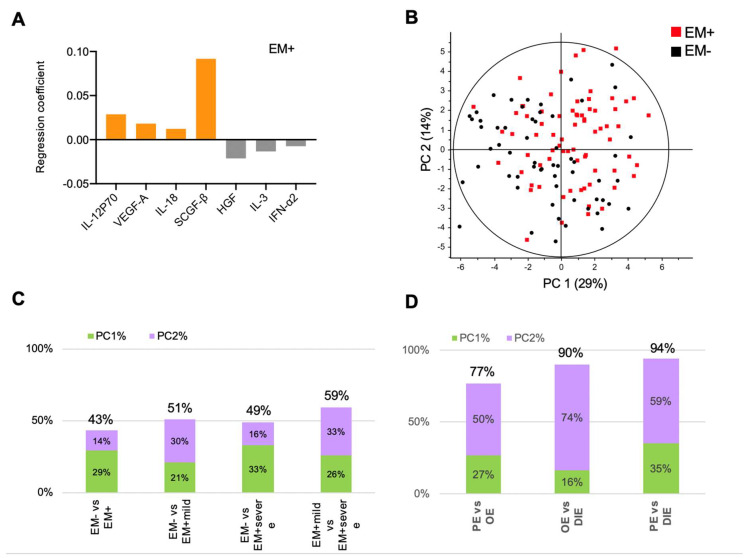
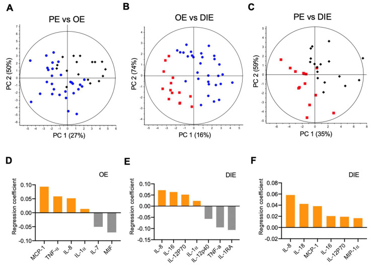
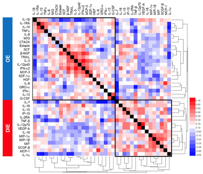
Endometriosis is a common inflammatory gynecological disorder which causes pelvic scarring, pain, and infertility, characterized by the implantation of endometrial-like lesions outside the uterus. The peritoneum, ovaries, and deep soft tissues are the commonly involved sites, and endometriotic lesions can be classified into three subphenotypes: superficial peritoneal endometriosis (PE), ovarian endometrioma (OE), and deep infiltrating endometriosis (DIE). In 132 women diagnosed laparoscopically with and without endometriosis (n = 73, 59 respectively), and stratified into PE, OE, and DIE, peritoneal fluids (PF) were characterized for 48 cytokines by using multiplex immunoassays. Partial-least-squares-regression analysis revealed distinct subphenotype cytokine signatures-a six-cytokine signature distinguishing PE from OE, a seven-cytokine signature distinguishing OE from DIE, and a six-cytokine-signature distinguishing PE from DIE-each associated with different patterns of biological processes, signaling events, and immunology. These signatures describe endometriosis better than disease stages (p < 0.0001). Pathway analysis revealed the association of ERK1 and 2, AKT, MAPK, and STAT4 linked to angiogenesis, cell proliferation, migration, and inflammation in the subphenotypes. These data shed new insights on the pathophysiology of endometriosis subphenotypes, with the potential to exploit the cytokine signatures to stratify endometriosis patients for targeted therapies and biomarker discovery.

Keywords: cytokines; endometriosis; microenvironment; peritoneal fluid; precision medicine.


Conflict of interest statement

The authors declare no conflict of interest.

Figures

Figure 1
Figure 1
Peritoneal fluid cytokines associate with endometriosis stages. (A) Partial least squares regression (PLSR) coefficient analysis revealed a signature comprising elevated IL-12p70, IL-18, VEGF-A, and SCGF-β and decreased IFN-α2, IL-3, and HGF that distinguished women with endometriosis (EM+) from women without (EM-). (B) Modeling by PLSR scores plot reveals overlap of EM- and EM+, suggesting heterogeneity in the peritoneal fluid environment. PLSR-derived principal component scores of principal component 1 (PC1) and principal component 2 (PC2) of (C) stages and (D) subphenotypes. Cumulative principal-component scores are shown at the top of each bar.
Figure 2
Figure 2
Peritoneal fluid cytokines show distinct delineation of endometriosis subphenotypes. Partial least squares regression (PLSR) models separated (A) ovarian endometriomas from peritoneal endometriosis, (B) ovarian endometriomas from deep infiltrating endometriosis, and (C) peritoneal endometriosis from deep infiltrating endometriosis. The principal component (PC) scores show good separation of endometriosis subphenotypes by using PF cytokines. (DF). Corresponding PLSR coefficient analyses reveal cytokine signatures delineating the various subphenotypes. Elevated cytokines associated with a particular endometriosis subphenotype (OE, PE, or DIE) relative to its comparator appear in the same upper or lower half of the plot.
Figure 3
Figure 3
Correlation matrix of peritoneal fluid cytokines. Hierarchical clustering was performed on Spearman r-values between the subphenotypes ovarian endometriosis, and deep infiltrating endometriosis revealed consistency in PLSR-derived cytokine signatures that segregated OE from DIE.

References

    1. Practice Committee of the American Society for Reproductive Medicine Endometriosis and infertility. Fertil. Steril. 2006;86:S156–S160. doi: 10.1016/j.fertnstert.2006.08.014. - DOI - PubMed
    1. Bulun S.E. Endometriosis. N. Engl. J. Med. 2009;360:268–279. doi: 10.1056/NEJMra0804690. - DOI - PubMed
    1. Nisolle M., Donnez J. Peritoneal endometriosis, ovarian endometriosis, and adenomyotic nodules of the rectovaginal septum are three different entities. Fertil. Steril. 1997;68:585–596. doi: 10.1016/S0015-0282(97)00191-X. - DOI - PubMed
    1. Stegmann B.J., Sinaii N., Liu S., Segars J., Merino M., Nieman L.K., Stratton P. Using location, color, size, and depth to characterize and identify endometriosis lesions in a cohort of 133 women. Fertil. Steril. 2008;89:1632–1636. doi: 10.1016/j.fertnstert.2007.05.042. - DOI - PMC - PubMed
    1. Burney R.O., Giudice L.C. Pathogenesis and pathophysiology of endometriosis. Fertil. Steril. 2012;98:511–519. doi: 10.1016/j.fertnstert.2012.06.029. - DOI - PMC - PubMed