. 2020 May 15;12(5):1254.
doi: 10.3390/cancers12051254.

Characteristics of MGUS and Multiple Myeloma According to the Target of Monoclonal Immunoglobulins, Glucosylsphingosine, or Epstein-Barr Virus EBNA-1

Affiliations

Characteristics of MGUS and Multiple Myeloma According to the Target of Monoclonal Immunoglobulins, Glucosylsphingosine, or Epstein-Barr Virus EBNA-1

Adrien Bosseboeuf et al. Cancers (Basel). .

Abstract

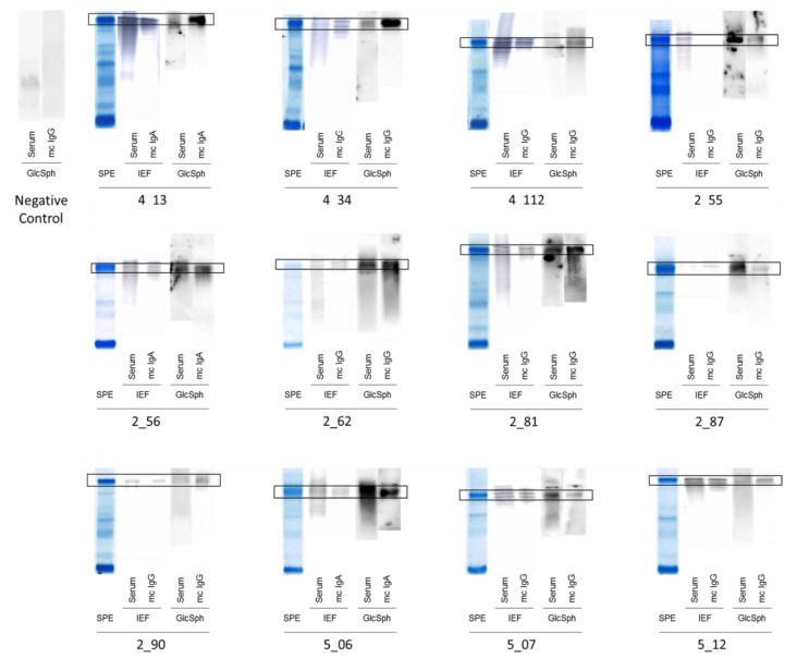
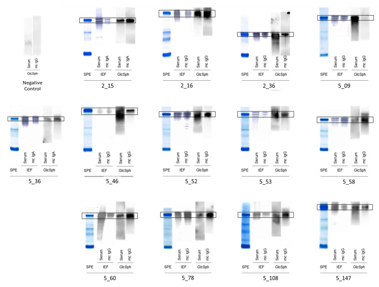
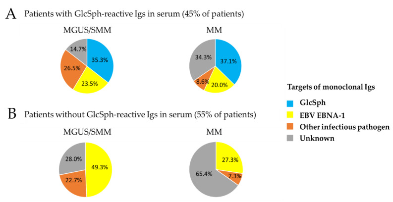
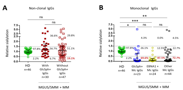
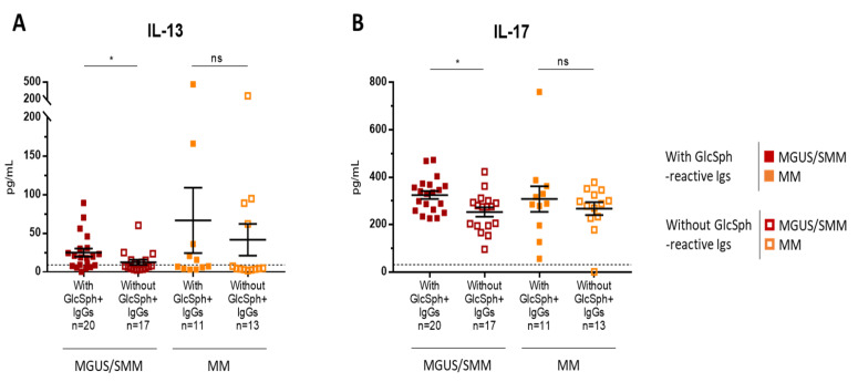
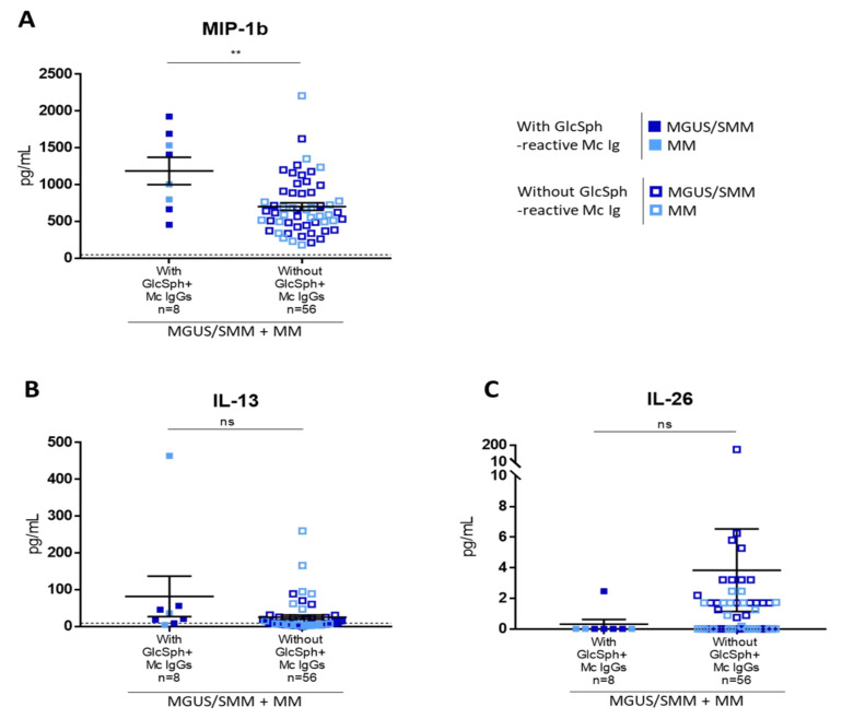
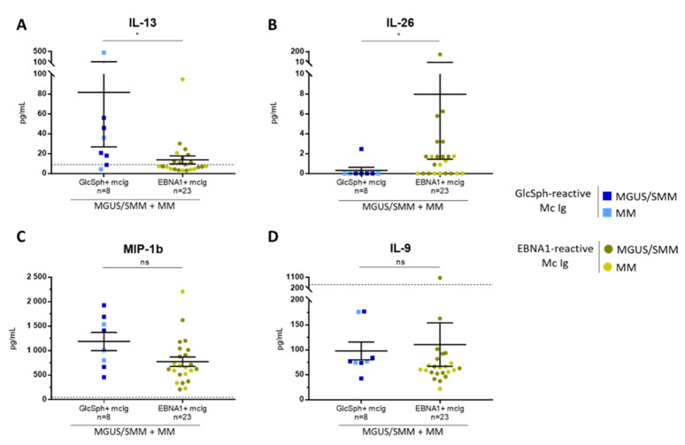
: Chronic stimulation by infectious or self-antigens initiates subsets of monoclonal gammopathies of undetermined significance (MGUS), smoldering multiple myeloma (SMM), or multiple myeloma (MM). Recently, glucosylsphingosine (GlcSph) was reported to be the target of one third of monoclonal immunoglobulins (Igs). In this study of 233 patients (137 MGUS, 6 SMM, 90 MM), we analyzed the GlcSph-reactivity of monoclonal Igs and non-clonal Igs. The presence of GlcSph-reactive Igs in serum was unexpectedly frequent, detected for 103/233 (44.2%) patients. However, GlcSph was targeted by the patient's monoclonal Ig for only 37 patients (15.9%); for other patients (44 MGUS, 22 MM), the GlcSph-reactive Igs were non-clonal. Then, the characteristics of patients were examined: compared to MM with an Epstein-Barr virus EBNA-1-reactive monoclonal Ig, MM patients with a GlcSph-reactive monoclonal Ig had a mild presentation. The inflammation profiles of patients were similar except for moderately elevated levels of 4 cytokines for patients with GlcSph-reactive Igs. In summary, our study highlights the importance of analyzing clonal Igs separately from non-clonal Igs and shows that, if autoimmune responses to GlcSph are frequent in MGUS/SMM and MM, GlcSph presumably represents the initial pathogenic event for ~16% cases. Importantly, GlcSph-initiated MM appears to be a mild form of MM disease.

Keywords: antigen specificity; auto-antigen; cytokine; glucosylsphingosine (GlcSph); inflammation; interleukin-13 (IL-13); interleukin-17 (IL-17); interleukin-26 (IL-26); lysoglucosyl-ceramide (LGL1); monoclonal gammopathy of undetermined significance (MGUS); monoclonal immunoglobulin; multiple myeloma; sialylation.


Conflict of interest statement

The authors declare that they have no conflict of interest and nothing to disclose.

Figures

Figure 1
Figure 1
Glucosylsphingosine (GlcSph)-reactivity of serum Igs and monoclonal Igs obtained for patients with monoclonal gammopathy of undetermined significance (MGUS). Measurement of Ig concentration, separation of monoclonal Igs from other Igs, and verification of purity were performed as published [5,6,7,14,15]. Purification starts with the separation of serum proteins with high resolution agarose gel electrophoresis (SAS-MX high resolution, Helena Biosciences, Gateshead, UK). Then, the monoclonal Ig is cut from the gel and eluted in phosphate buffer saline (PBS). The purity of the monoclonal Ig preparation is verified by isoelectric focusing (IEF) on an agarose gel (pH 3–10) followed by blotting and immuno-revelation by an anti-human IgGγ chain or anti-IgAα chain antibody labeled with peroxidase. For GlcSph-specific immunoblotting, polyvinylidene fluoride (PVDF) membranes are incubated for 90 min in 100 μg/mL of GlcSph in 0.1 M sodium bicarbonate, rinsed in PBS and 0.1% Tween 20 detergent, and then blocked with 5% bovine serum albumin (BSA) in PBS and 0.1% Tween 20. Samples of serum and purified monoclonal Ig are submitted to agarose gel electrophoresis, and then the gels are blotted onto the GlcSph-saturated membranes by diffusion blotting during 12 min. After blocking with 2.5% BSA in PBS and 0.1% Tween 20, membranes are incubated with anti-human IgG or IgA horseradish peroxidase (HRP)-conjugated secondary antibody, washed, and revealed by chemiluminescence. Signals corresponding to the patient’s monoclonal Ig are encircled in black. The negative control is a patient with no GlcSph-reactive Ig in serum. SPE = Serum protein electrophoresis; Mc Ig = purified monoclonal Ig. The GlcSph-reactivity of serum Igs and monoclonal Igs obtained for 12 other MGUS patients are shown in Figure S2.
Figure 2
Figure 2
GlcSph-reactivity of serum Igs and monoclonal Igs in MM. Measurement of Ig concentration, separation of monoclonal Igs from other Igs, and verification of purity are performed as described above (Figure 1), in Methods, and published previously [5,6,7,14,15]. SPE = Serum protein electrophoresis; Mc Ig = purified monoclonal Ig. Signals corresponding to the patient’s monoclonal Ig are encircled. The negative control is a patient with no GlcSph-reactive Ig in serum.
Figure 3
Figure 3
Identified targets of monoclonal Igs in MGUS/SMM and MM, according to the presence or absence of GlcSph-reactive Igs. Representation of the percentage (%) of patients with a monoclonal Ig that targets GlcSph, EBV EBNA-1, or another MIAA pathogen. “Unknown”: patients for whom the target of the monoclonal Ig has not been identified (unknown target). (A) Targets of monoclonal Igs from patients with GlcSph-reactive Igs (clonal or non-clonal). (B) Targets of monoclonal Igs from patients who do not have GlcSph-reactive Igs.
Figure 4
Figure 4
Level of sialylation of IgGs from MGUS/SMM and MM patients. Sialylation studies were performed using an ELLA (Enzyme Linked Lectin Assay) for IgG sialylation detection, as well as an ELISA (Enzyme Linked Immuno Sorbent Assay) for total IgG detection [15]. Clonal and non-clonal IgG fractions were prepared as described in Methods and published [15]. The relative sialylation of IgGs was expressed as the sialic acid/IgG optic density (OD) ratio. (A) Degree of sialylation of polyclonal IgGs from 43 healthy donors (HD) (OD ratio between 0.5 and 1.5) and of non-clonal IgGs from 77 patients. (B) Degree of sialylation of 91 purified monoclonal IgGs analyzed according to the target of the monoclonal IgG. Bars indicate means ± SEM. The percentages indicate the % of IgGs with a low (OD ratio < 0.5), normal (OD ratio: 0.5–1.5) or high (OD ratio > 0.5) degree of sialylation. Statistical analysis was performed using one-way ANOVA test followed by Tukey’s multiple comparison test. * p < 0.05, ** p < 0.01, *** p < 0.001. NS: not significant.
Figure 5
Figure 5
Differences in cytokine levels according to the presence of GlcSph-reactive polyclonal Ig(s) for MGUS/ SMM and MM patients. Forty-two cytokines, chemokines and receptors were quantified using the Luminex technology and Bio-Plex Pro Human Cytokine Panel kits [15]. The levels of only 2 cytokines, IL-13 (A) and IL-17 (B), were significantly different depending on the presence of polyclonal GlcSph-reactive IgGs. Bars indicate means + SEM; note that each figure has a different scale. GlcSph+: with GlcSph-reactive IgG(s) in serum. MGUS/SMM patients with () or without () GlcSph-reactive IgGs in serum; MM patients with () or without () GlcSph-reactive IgGs in serum. (*) p < 0.05, Mann–Whitney t-test. The dotted line represents the maximal normal value observed in healthy individuals. NS: not significant.
Figure 6
Figure 6
Differences in cytokine levels according to the presence of a GlcSph-reactive monoclonal Ig for MGUS/SMM and MM patients. The levels of MIP-1β (A) were significantly different depending on the presence of a monoclonal GlcSph-reactive IgG, whereas the levels of IL-13 (B) and IL-26 (C) were not. Each figure has a different scale. Bars indicate means + SEM. MGUS/SMM () and MM () patients with a GlcSph-reactive monoclonal (Mc) IgG; MGUS/SMM () and MM () patients without a GlcSph-reactive Mc IgG. (**) p < 0.01, Mann–Whitney t-test. The dotted line represents the maximal normal value observed in healthy individuals. NS: not significant.
Figure 7
Figure 7
Differences in cytokine levels according to the presence of GlcSph- or EBV EBNA-1-reactive monoclonal Ig for MGUS/SMM and MM patients. The levels of 2 cytokines, IL-13 (A) and IL-26 (B) were significantly different depending on the antigen specificity of the monoclonal Ig, whereas the levels of MIP-1β (C) and IL-9 (D) were not. Note that each figure has a different scale; bars indicate means ± SEM. GlcSph+: with GlcSph-reactive monoclonal (mc) IgG; EBNA-1+: with EBV EBNA-1-reactive mc IgG. () MGUS/SMM and () MM patients with GlcSph-reactive mc IgG; () MGUS/SMM and () MM patients with an EBNA-1-reactive mc IgG. (*) p < 0.05, Mann–Whitney t-test. The dotted line represents the highest normal value observed in healthy donors. NS: not significant.

Similar articles

Cited by

References

    1. Dhodapkar M.V. MGUS to myeloma: A mysterious gammopathy of underexplored significance. Blood. 2016;128:2599–2606. doi: 10.1182/blood-2016-09-692954. - DOI - PMC - PubMed
    1. Kyle R.A., Larson D.R., Therneau T.M., Dispenzieri A., Kumar S., Cerhan J.R., Rajkumar S.V. Long-Term Follow-up of Monoclonal Gammopathy of Undetermined Significance. N. Engl. J. Med. 2018;378:241–249. doi: 10.1056/NEJMoa1709974. - DOI - PMC - PubMed
    1. Boyle E.M., Davies F.E., Leleu X., Morgan G.J. Understanding the multiple biological aspects leading to myeloma. Haematologica. 2014;99:605–612. doi: 10.3324/haematol.2013.097907. - DOI - PMC - PubMed
    1. Rajkumar S.V., Dimopoulos M.A., Palumbo A., Blade J., Merlini G., Mateos M.V., Kumar S., Hillengass J., Kastritis E., Richardson P., et al. International Myeloma Working Group updated criteria for the diagnosis of multiple myeloma. Lancet Oncol. 2014;15:e538–e548. doi: 10.1016/S1470-2045(14)70442-5. - DOI - PubMed
    1. Bosseboeuf A., Feron D., Tallet A., Rossi C., Charlier C., Garderet L., Caillot D., Moreau P., Cardo-Vila M., Pasqualini R., et al. Monoclonal IgG in MGUS and multiple myeloma target infectious pathogens. J. Clin. Investig. Insight. 2017;2:95367. doi: 10.1172/jci.insight.95367. - DOI - PMC - PubMed

LinkOut - more resources