

Multi-Level Cross Residual Network for Lung Nodule Classification
- PMID: 32429401
- PMCID: PMC7284728
- DOI: 10.3390/s20102837
Multi-Level Cross Residual Network for Lung Nodule Classification
Abstract
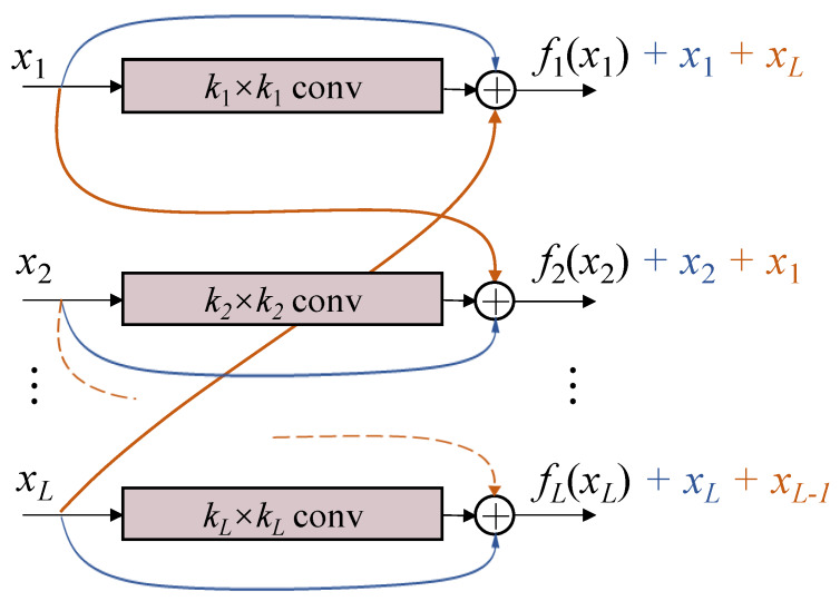
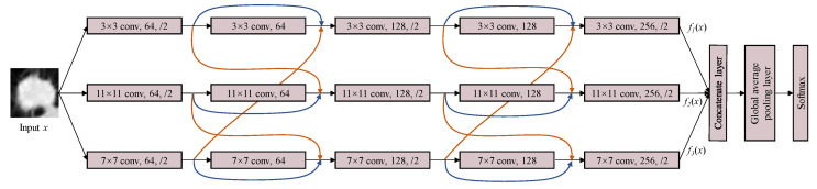
Computer-aided algorithm plays an important role in disease diagnosis through medical images. As one of the major cancers, lung cancer is commonly detected by computer tomography. To increase the survival rate of lung cancer patients, an early-stage diagnosis is necessary. In this paper, we propose a new structure, multi-level cross residual convolutional neural network (ML-xResNet), to classify the different types of lung nodule malignancies. ML-xResNet is constructed by three-level parallel ResNets with different convolution kernel sizes to extract multi-scale features of the inputs. Moreover, the residuals are connected not only with the current level but also with other levels in a crossover manner. To illustrate the performance of ML-xResNet, we apply the model to process ternary classification (benign, indeterminate, and malignant lung nodules) and binary classification (benign and malignant lung nodules) of lung nodules, respectively. Based on the experiment results, the proposed ML-xResNet achieves the best results of 85.88% accuracy for ternary classification and 92.19% accuracy for binary classification, without any additional handcrafted preprocessing algorithm.
Keywords: binary; computed tomography; lung nodule classification; residual convolutional neural network; ternary.
Conflict of interest statement
The authors declare no conflict of interest.
Figures
References
-
- Ferlay J., Ervik M., Lam F., Colombet M., Mery L., Pi neros M., Znaor A., Soerjomataram I., Bray F. Global Cancer Observatory: Cancer Today. International Agency for Research on Cancer; Lyon, France: 2018.
MeSH terms
LinkOut - more resources
Full Text Sources
Medical
Research Materials
