. 2020 May 16;9(5):1237.
doi: 10.3390/cells9051237.

Exogenous Liposomal Ceramide-C6 Ameliorates Lipidomic Profile, Energy Homeostasis, and Anti-Oxidant Systems in NASH

Affiliations

Exogenous Liposomal Ceramide-C6 Ameliorates Lipidomic Profile, Energy Homeostasis, and Anti-Oxidant Systems in NASH

Francesca Zanieri et al. Cells. .

Abstract

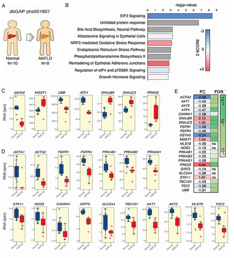
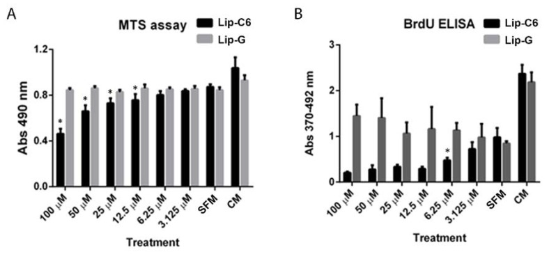
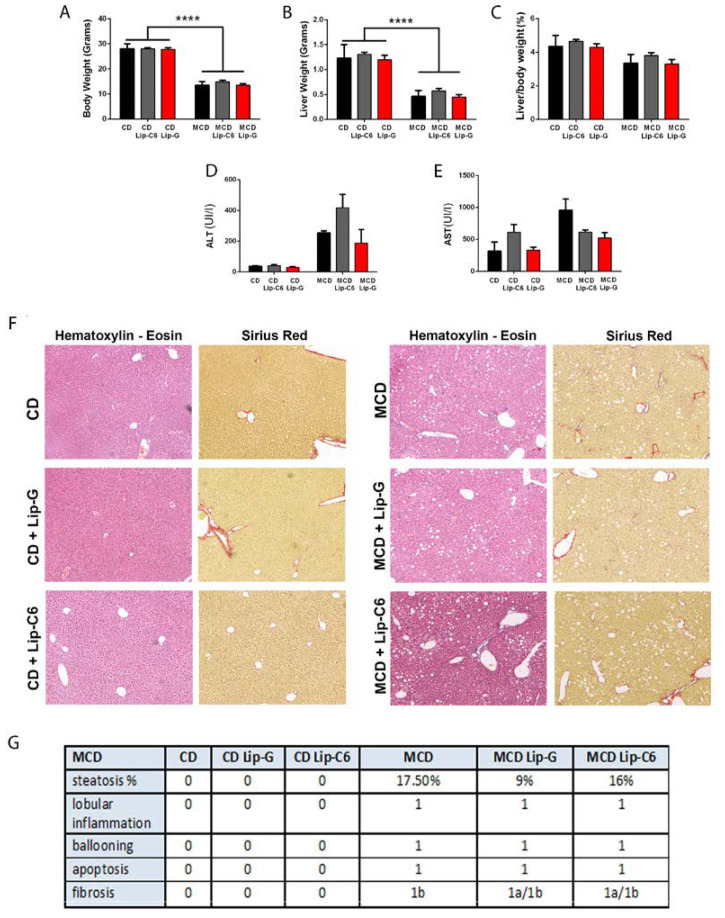
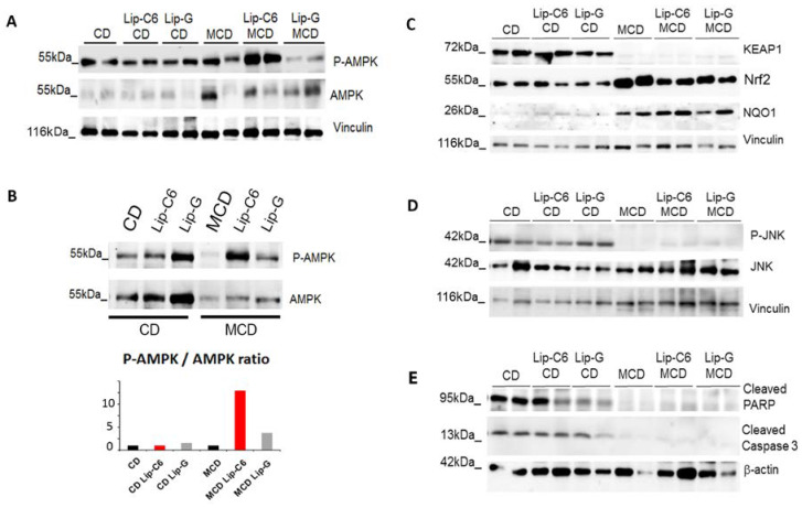
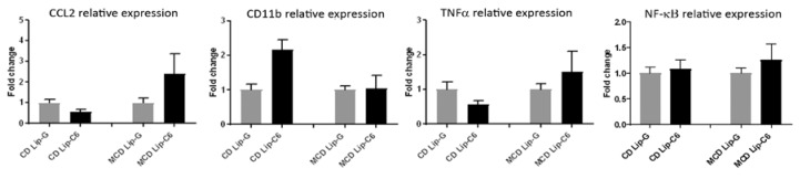
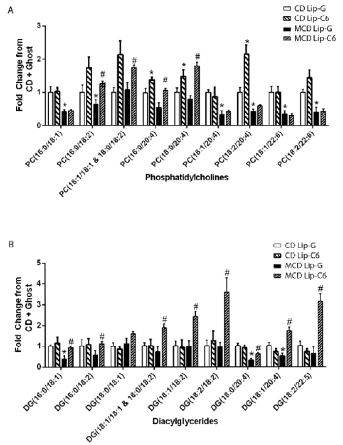
In non-alcoholic steatohepatitis (NASH), many lines of investigation have reported a dysregulation in lipid homeostasis, leading to intrahepatic lipid accumulation. Recently, the role of dysfunctional sphingolipid metabolism has also been proposed. Human and animal models of NASH have been associated with elevated levels of long chain ceramides and pro-apoptotic sphingolipid metabolites, implicated in regulating fatty acid oxidation and inflammation. Importantly, inhibition of de novo ceramide biosynthesis or knock-down of ceramide synthases reverse some of the pathology of NASH. In contrast, cell permeable, short chain ceramides have shown anti-inflammatory actions in multiple models of inflammatory disease. Here, we investigated non-apoptotic doses of a liposome containing short chain C6-Ceramide (Lip-C6) administered to human hepatic stellate cells (hHSC), a key effector of hepatic fibrogenesis, and an animal model characterized by inflammation and elevated liver fat content. On the basis of the results from unbiased liver transcriptomic studies from non-alcoholic fatty liver disease patients, we chose to focus on adenosine monophosphate activated kinase (AMPK) and nuclear factor-erythroid 2-related factor (Nrf2) signaling pathways, which showed an abnormal profile. Lip-C6 administration inhibited hHSC proliferation while improving anti-oxidant protection and energy homeostasis, as indicated by upregulation of Nrf2, activation of AMPK and an increase in ATP. To confirm these in vitro data, we investigated the effect of a single tail-vein injection of Lip-C6 in the methionine-choline deficient (MCD) diet mouse model. Lip-C6, but not control liposomes, upregulated phospho-AMPK, without inducing liver toxicity, apoptosis, or exacerbating inflammatory signaling pathways. Alluding to mechanism, mass spectrometry lipidomics showed that Lip-C6-treatment reversed the imbalance in hepatic phosphatidylcholines and diacylglycerides species induced by the MCD-fed diet. These results reveal that short-term Lip-C6 administration reverses energy/metabolic depletion and increases protective anti-oxidant signaling pathways, possibly by restoring homeostatic lipid function in a model of liver inflammation with fat accumulation.

Keywords: adenosine monophosphate-activated kinase (AMPK); apoptosis; ceramides; diacylglycerol (DG); human hepatic stellate cells (hHSC); inflammation; lipidomics; liposomes; methionine-choline deficient diet (MCD); non-alcoholic steatohepatitis (NASH); nuclear factor-erythroid 2-related factor 2 (Nfe2l2/NRF2); phosphatidylcholine (PC).


Conflict of interest statement

“The funders had no role in the design of the study; in the collection, analyses, or interpretation of data; in the writing of the manuscript; or in the decision to publish the results”. Penn State Research Foundation has licensed ceramide nanotechnology to Keystone Nano, Inc. (PA, USA) and M.K. is cofounder and Chief Medical Officer of Keystone Nano. R.B. has done consulting services for Verlyx Pharma Inc. G.M., L.F., and L.L. are now full time employees at Engitix Ltd., and G.M., L.L., M.P., and K.R. own shares in Engitix Ltd. M.P. and K.R. receive consultancies from Engitix Ltd.

Figures

Figure 1
Figure 1
Adenosine monophosphate-activated kinase (AMPK) subunits and Nrf2 gene expression are significantly changed in nonalcoholic fatty liver disease (NAFLD) patients. RNA sequencing was performed on (A) normal (N = 10) and NAFLD derived liver tissue (N = 9 patients). (B) Summary of the functional enrichment analysis of the differentially expressed transcriptome using ingenuity pathway analysis. The significance of each gene set is indicated in the bar graph. The top functions belonging to the canonical pathway class are represented. The colors of the bars indicate the direction (blue meaning inhibition, and red activation) and the Z-score, which measures the magnitude (intensity of color) for the enrichment of a specific pathway. For some functions, no Z-score was calculated because of a lack of enough evidence regarding the specific gene functions or expression within a specific gene set (gray bars). The pathway “NRF2-mediated oxidative stress response” was significantly upregulated in patients with NAFLD. (CD) Gene expression related to different Nrf2 and AMPK signaling pathways/mechanisms shown to be altered. (E) Representations of fold change (FC) and false discovery ratio (FDR) are shown for each gene investigated.
Figure 2
Figure 2
Liposomes containing short-chain ceramide C6 affect AMPK activation and proliferation, and promote the endogenous anti-oxidant system in primary human HSCs. Primary hHSCs were exposed to various concentrations (100–3.125 µM) of Lip-C6 or Lip-G for up to 24 h. (A) Higher doses of Lip-C6-treatment induced cytotoxicity (100–12.5 µM) (n = 4 per condition) (* p < 0.05), whereas (B) 6.25 µM inhibited hHSC proliferation (* p < 0.05) compared with serum free medium (SFM) (n = 4 per condition). (C) Representative images of liposomal uptake, which were evaluated by employing immunofluorescence and showed that liposomes containing ceramide-C6 with rhodamine (6.25 µM) were taken up as early as two hours after exposure. (D) Primary hHSCs were exposed to 6.25 and 3.125 µM of Lip-C6 for up to 24 h. Representative protein analysis showed an increase in phosphorylation of AMPK and upregulation of Nrf2 protein expression (n = 1 of 3 independent experiments). (E) Changes in ATP were observed when cells were exposed to 6.25 µM of Lip-C6 for up to 24 h (n = 4 per condition, * p < 0.05, ** p < 0.005 compared with SFM), Complete Medium (CM).
Figure 2
Figure 2
Liposomes containing short-chain ceramide C6 affect AMPK activation and proliferation, and promote the endogenous anti-oxidant system in primary human HSCs. Primary hHSCs were exposed to various concentrations (100–3.125 µM) of Lip-C6 or Lip-G for up to 24 h. (A) Higher doses of Lip-C6-treatment induced cytotoxicity (100–12.5 µM) (n = 4 per condition) (* p < 0.05), whereas (B) 6.25 µM inhibited hHSC proliferation (* p < 0.05) compared with serum free medium (SFM) (n = 4 per condition). (C) Representative images of liposomal uptake, which were evaluated by employing immunofluorescence and showed that liposomes containing ceramide-C6 with rhodamine (6.25 µM) were taken up as early as two hours after exposure. (D) Primary hHSCs were exposed to 6.25 and 3.125 µM of Lip-C6 for up to 24 h. Representative protein analysis showed an increase in phosphorylation of AMPK and upregulation of Nrf2 protein expression (n = 1 of 3 independent experiments). (E) Changes in ATP were observed when cells were exposed to 6.25 µM of Lip-C6 for up to 24 h (n = 4 per condition, * p < 0.05, ** p < 0.005 compared with SFM), Complete Medium (CM).
Figure 3
Figure 3
Liposomal treatment with ceramide-C6 in methionine-choline deficient (MCD)-induced liver steatosis. (A) Animals fed the MCD diet have significant loss of body weight regardless of treatment with Lip-C6 or Lip-G in comparison with the control diet (CD) group (**** p < 0.0001). (B) This coincided with a strong significant decrease in liver size in comparison with the control diet (CD) group (**** p < 0.0001) without (C) changes in liver/body weight ratio when comparing the control diet with MCD-fed mice. (D,E) No significant differences were observed between alanine transaminase (ALT) and aspartate aminotransferase (AST) levels of animals treated with Lip-C6 or Lip-G in comparison with their specific control condition. (F) Lip-C6 treatment did not exacerbate the MCD diet, as analyzed by hematoxylin and eosin and Sirius Red. (G) NASH CRN scoring system demonstrating changes occurring during Lip-G and Lip-C6 treatment in MCD-fed mice (CD n = 5; CD–Lip-G n = 5; CD–Lip-C6 n = 5; MCD n = 5; MCD Lip-G n = 5; and MCD–Lip-C6 n = 9).
Figure 4
Figure 4
Liposome containing short-chain ceramide C6 treatment in MCD diet increases AMPK activation/phosphorylation and enhances the endogenous anti-oxidative stress signaling pathway without inducing apoptosis. (A) Representative Western blot analysis demonstrated an upregulation in activation/phosphorylation of AMPK by Lip-C6 treatment in MCD-fed mice in comparison with MCD-fed mice (n = 2 for each condition). (B) Inter-variability was assessed by protein analysis of pooled samples of each condition and densitometry scanning. MCD-fed mice have reduced AMPK protein levels in comparison with control diet-fed mice. Absolute levels of phosphorylated AMPK (P-AMPK) were highly induced in MCD-fed mice treated with Lip-C6 relative to MCD-fed mice and MCD-fed Lip-G treated mice. (C) Representative Western blot analysis demonstrated that Keap-1 protein expression was absent in MCD-fed mice, whereas Nrf2 and NQO1 protein expression were upregulated in MCD-fed mice in comparison with CD-fed mice (n = 2 for each condition). (D) Lip-C6 treatment in MCD-fed mice did not induce apoptosis by JNK activation/phosphorylation. (E) Cleaved poly (ADP-ribose) polymerase (PARP) and cleaved Caspase 3 protein expression showed to be absent in MCD-fed mice with no changes observed in Lip-C6 treated mice (n = 2 for each condition).
Figure 5
Figure 5
Lip-C6 does not exacerbate the pro-inflammatory response in MCD-fed mice. qRT-PCR showed that the Lip-C6 treatment in MCD-fed mice did not induce significant changes in mRNA expression of pro-inflammatory key genes versus internal control diet (CD) (CD n = 2, CD-G n = 5, CD-C6 n = 5, MCD n = 2, MCD-G n = 5, and MCD-C6 n = 8, mean value of controls was set to 1).
Figure 6
Figure 6
Lip-C6 treatment restores specific phosphatidylcholines (PC) and diacylglycerides (DC) in MCD-fed mice. Animals received MCD or CD diet for nine weeks and were further subdivided and administered a single tail vein injection of Lip-C6 or Lip-G. One week after treatment, all mice were euthanized. Lipids from liver samples were extracted and untargeted LC-MS/MS lipidomics approach was performed. (A) Major changes were observed in the lipid classes of phosphatidylcholine (PC) and (B) diacylglycerol classes (DG) when MCD-fed mice treated with Lip-C6 were compared with MCD-fed mice treated with Lip-G. No changes were detected for short-chain DG and TG. * p < 0.05 CMCD + Lip-G (n = 5) vs. MCD + Lip-G (n = 5), CMCD + Lip-G vs. CMCD + Lip-C6 (n = 8) # p < 0.05 MCD + Lip-G vs. MCD + Lip-C6 (n = 9).

Similar articles

Cited by

References

    1. Blachier M., Leleu H., Peck-Radosavljevic M., Valla D.C., Roudot-Thoraval F. The burden of liver disease in Europe: A review of available epidemiological data. J. Hepatol. 2013;58:593–608. doi: 10.1016/j.jhep.2012.12.005. - DOI - PubMed
    1. Yeh M.M., Brunt E.M. Pathological features of fatty liver disease. Gastroenterology. 2014;147:754–764. doi: 10.1053/j.gastro.2014.07.056. - DOI - PubMed
    1. Bataller R., Rombouts K., Altamirano J., Marra F. Fibrosis in alcoholic and nonalcoholic steatohepatitis. Best. Pract. Res. Clin. Gastroenterol. 2011;25:231–244. doi: 10.1016/j.bpg.2011.02.010. - DOI - PubMed
    1. Drescher H.K., Weiskirchen S., Weiskirchen R. Current Status in Testing for Nonalcoholic Fatty Liver Disease (NAFLD) and Nonalcoholic Steatohepatitis (NASH) Cells. 2019;8:845. doi: 10.3390/cells8080845. - DOI - PMC - PubMed
    1. Noureddin M., Sanyal A.J. Pathogenesis of NASH: The Impact of Multiple Pathways. Curr. Hepatol. Rep. 2018;17:350–360. doi: 10.1007/s11901-018-0425-7. - DOI - PMC - PubMed

Publication types

MeSH terms