. 2020 May 16;9(5):1238.
doi: 10.3390/cells9051238.

Functional Characterization of Neurofilament Light Splicing and Misbalance in Zebrafish

Affiliations

Functional Characterization of Neurofilament Light Splicing and Misbalance in Zebrafish

Doris Lou Demy et al. Cells. .

Abstract

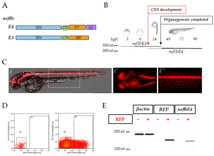
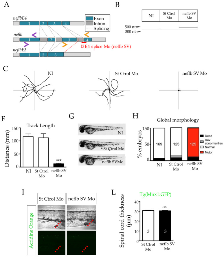
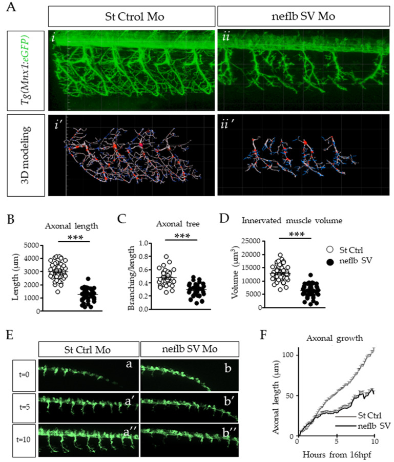
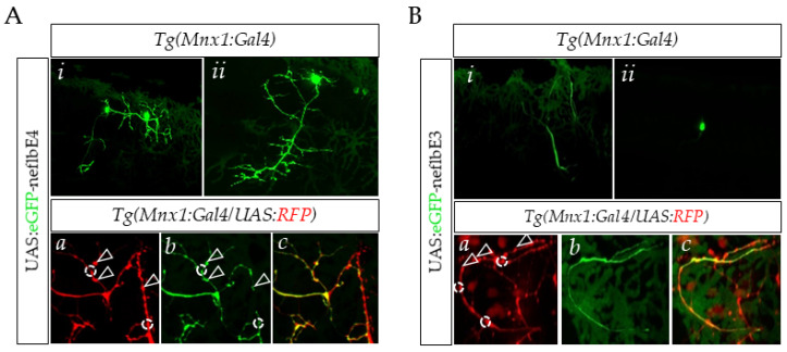
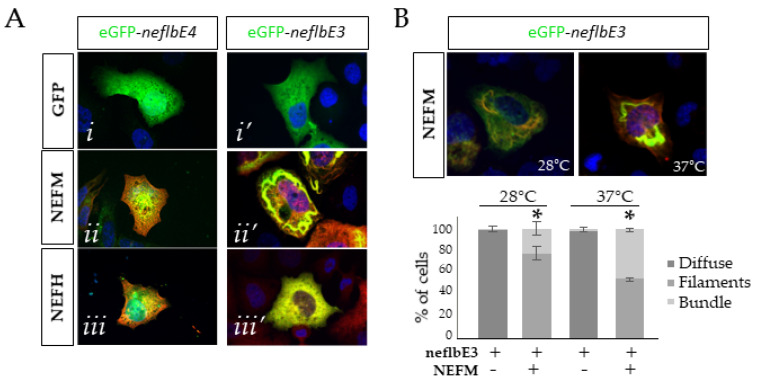
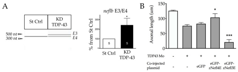
Neurofilaments (NFs), a major cytoskeletal component of motor neurons, play a key role in the differentiation, establishment and maintenance of their morphology and mechanical strength. The de novo assembly of these neuronal intermediate filaments requires the presence of the neurofilament light subunit (NEFL), whose expression is reduced in motor neurons in amyotrophic lateral sclerosis (ALS). This study used zebrafish as a model to characterize the NEFL homologue neflb, which encodes two different isoforms via a splicing of the primary transcript (neflbE4 and neflbE3). In vivo imaging showed that neflb is crucial for proper neuronal development, and that disrupting the balance between its two isoforms specifically affects the NF assembly and motor axon growth, with resultant motor deficits. This equilibrium is also disrupted upon the partial depletion of TDP-43 (TAR DNA-binding protein 43), an RNA-binding protein encoded by the gene TARDBP that is mislocalized into cytoplasmic inclusions in ALS. The study supports the interaction of the NEFL expression and splicing with TDP-43 in a common pathway, both biologically and pathogenetically.

Keywords: amyotrophic lateral sclerosis (ALS), neurofilament light (NEFL), TDP-43; neurofilaments (NFs); zebrafish.


Conflict of interest statement

The authors declare no conflict of interest.

Figures

Figure 1
Figure 1
neflbE3 and neflbE4 expression in zebrafish. (A), schematic of the zebrafish exon structures of the predicted neflb splice variants. Length (bp) is indicated on each exon. (B), neflb is expressed at all embryonic and larval stages in zebrafish, with a splicing shift from neflb3E (upper PCR band) to neflb4E (lower PCR band) occurring during CNS development—revealed by a change in the amplicon size. (C) (i–i″), sagittal and dorsal vision of Tg(HuC:Gal4/UAS:RFP) zebrafish embryos at 48hpf. This transgenic line expresses the red fluorescent protein (RFP) in post-mitotic neurons. (D), post-mitotic neurons (RFP+) and non-neuronal cells (RFP-) were isolated by FACS, and the expression of β-actin, RFP and neflb4E was tested by PCR in both pools (E).
Figure 2
Figure 2
neflbE3/E4 misbalance results in a strong and specific motor phenotype. (A), position of the splicing variant (SV) antisense oligomorpholino (neflb SV Mo, in red) targeting the decisive I3E4 splice junction in order to inhibit the developmental shift from neflb3E to neflb4E, and of the primer pairs used in this study: ZF Neflb (purple pair) and ZF Neflb SV (orange pair). (B), St Ctrol Mo and neflb SV Mo were injected at the dose of 0.2 mM. The I3E4 morpholino was efficient and caused the persistence of the neflb3E (upper band) expression at 48 hpf, when only neflb4E (lower band) was expressed in controls at this stage. (C), trajectories of 9 representative zebrafish embryos per condition during the touch-evoked escape response at 48 hpf. Non-injected (NI) and control injected embryos (St Ctrol) swam to the edges of a Petri dish in reaction to a light touch; neflb SV morphants were unable to move away from the center of the dish. (F), quantification of the track length shows a reduction by 90% in the swimming distance in neflb SV Mo-injected fish compared with the controls. (G), neflb SV Mo-injected embryos develop without any major developmental abnormality. (H), bar graph of the phenotype distribution after the TEER analysis at 48 hpf. Percentages of zebrafish with motor deficits (motor) are increased after the neflb SV Mo injection. Percentages of normally developed embryos in NI and St Ctrl Mo conditions are comparable. (I), Acridine orange staining (green), which is a metachromatic intercalator sensitive to DNA conformation, is used in this study to detect apoptosis. No higher amount of acridine orange-positive cells was detected in the spinal cord of the neflb SV morphants (right panel) compared with the controls (left panel). The urogenital opening (red dotted lines) has been used as a geographical reference for the measures. (L), spinal cord thickness measured at 48 hpf. *** p < 0.001; ns, non-significant.
Figure 3
Figure 3
Axonal atrophy in motor neurons of neflb SV embryos. (A), in vivo observation of somitic nerve fascicles in 48 hpf Hb9:eGFP zebrafish embryos injected with a control Mo (i) or with neflb SV Mo (ii). Hb9:eGFP line express the GFP (green) in motor neurons under the specific promoter HB9. After the neflb SV Mo injection, the axonal projections appeared shorter and less regularly distributed and branched than controls. (i’,ii’), 3D modeling of motor neuron morphology using the Imaris software. This reconstruction permitted the quantitative analysis of the nerve fascicles features: (B), the axonal length; (C), the axonal branching; (D), the volume of the innervated muscle. All measurements were reduced in the neflb SV morphants (black circles) with respect to the controls (white circles). (E), in vivo time-lapse imaging of the Tg(Mnx1:eGFP) zebrafish embryos injected with a control morpholino (a–a″) or with the neflb SV morpholino (b–b″) was performed during 10 h, starting from 16 hpf. (F), quantification of motor neurons axonal length during time. hpf, hour post fertilization. *** p < 0.001.
Figure 4
Figure 4
neflbE4 and neflbE3 distribution within motor neurons. (A) (i,ii), in vivo observation of single motor neurons expressing eGFP-neflbE4. Motor neurons expressing eGFP-neflb4E extend long and ramified axons. (a–c), eGFP-neflbE4 is particularly abundant in the specific area representing the axonal branching (white dotted circles) and NMJ buttons (white arrows). (B) (i,ii), single motor neurons expressing eGFP-neflb3E. eGFP-neflbE3 mostly aggregated or assembled into a long, massive, unramified bundle. (a–c), eGFP-neflbE3 was reduced or absent in the axonal ramification sites and NMJ buttons.
Figure 5
Figure 5
The neflb splicing variants assembly properties in SW13vim cells. (A), eGFP-neflbE4 (i) and eGFP-neflbE3 (i’) were expressed in SW13vim cells alone or together with the mouse NEFM (ii,ii’, red) or NEFH (iii,iii’, red). The assembly pattern was characterized at 48 h post transfection at 37 °C. (B), eGFP-neflbE3 and NEFM were co-transfected and the bundle formation was observed at 37 °C and 28 °C. Bottom panel, bar graph showing the percentage of cells carrying filaments, bundles or diffuse labeling. At 28 °C, the proportion of cells carrying bundles made of neflb3E and NEFM was less than the proportion of cells carrying bundles at 37 °C. * p < 0.05, (n = 3 cultures, 60 to 100 cells per coverslip) one-way ANOVA Tukey’s HSD post-hoc analysis.
Figure 6
Figure 6
TDP-43 regulates neflb splicing in zebrafish. (A) left, PCR analysis on RNA extracted from St Ctrl embryos and fish KD for TDP-43, using specific primers for both isoforms neflbE4 and neflbE3. Right, quantification of the neflbE3 and neflbE4 ratio. (B), axonal length measurement at 48 hpf after the overexpression of eGFP-neflbE4 and eGFP-neflbE3 constructs in KD TDP-43 embryos. neflbE4 significantly rescued the axon length (middle histo bar), whereas neflbE3 significantly aggravated the KD TDP-43 phenotype (right histo bar). * p < 0.05, *** p < 0.001, in comparison to TDP-43 Mo + eGFP.

References

    1. Hoffman P.N., Cleveland D.W., Griffin J.W., Landes P.W., Cowan N.J., Price D.L. Neurofilament gene expression: A major determinant of axonal caliber. Proc. Natl. Acad. Sci. USA. 1987;84:3472–3476. doi: 10.1073/pnas.84.10.3472. - DOI - PMC - PubMed
    1. Yuan A., Rao M.V., Nixon R.A. Neurofilaments and neurofilament proteins in health and disease. Cold Spring Harb. Perspect. Biol. 2017;9:a018309. doi: 10.1101/cshperspect.a018309. - DOI - PMC - PubMed
    1. Khalil M., Teunissen C.E., Otto M., Piehl F., Sormani M.P., Gattringer T., Barro C., Kappos L., Comabella M., Fazekas F., et al. Neurofilaments as biomarkers in neurological disorders. Nat. Rev. Neurol. 2018;14:577–589. doi: 10.1038/s41582-018-0058-z. - DOI - PubMed
    1. Leung C.L., Liem R.K. Characterization of interactions between the neurofilament triplet proteins by the yeast two-hybrid system. J. Biol. Chem. 1996;271:14041–14044. doi: 10.1074/jbc.271.24.14041. - DOI - PubMed
    1. Scott D., Smith K.E., O’Brien B.J., Angelides K.J. Characterization of mammalian neurofilament triplet proteins. Subunit stoichiometry and morphology of native and reconstituted filaments. J. Biol. Chem. 1985;260:10736–10747. - PubMed

Publication types

MeSH terms

Grants and funding

LinkOut - more resources