Review
. 2020 May 20;27(1):64.
doi: 10.1186/s12929-020-00653-y.

Molecular networking in the neuronal ceroid lipofuscinoses: insights from mammalian models and the social amoeba Dictyostelium discoideum

Affiliations
Review

Molecular networking in the neuronal ceroid lipofuscinoses: insights from mammalian models and the social amoeba Dictyostelium discoideum

Robert J Huber. J Biomed Sci. .

Erratum in

Abstract

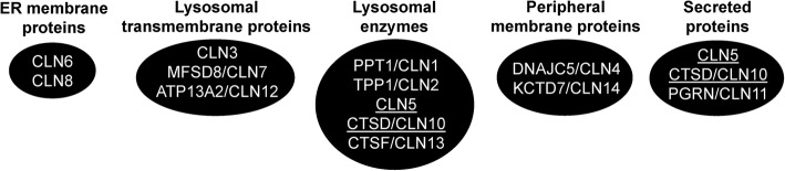
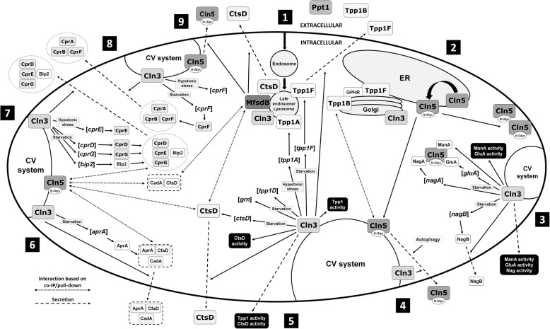
The neuronal ceroid lipofuscinoses (NCLs), commonly known as Batten disease, belong to a family of neurological disorders that cause blindness, seizures, loss of motor function and cognitive ability, and premature death. There are 13 different subtypes of NCL that are associated with mutations in 13 genetically distinct genes (CLN1-CLN8, CLN10-CLN14). Similar clinical and pathological profiles of the different NCL subtypes suggest that common disease mechanisms may be involved. As a result, there have been many efforts to determine how NCL proteins are connected at the cellular level. A main driving force for NCL research has been the utilization of mammalian and non-mammalian cellular models to study the mechanisms underlying the disease. One non-mammalian model that has provided significant insight into NCL protein function is the social amoeba Dictyostelium discoideum. Accumulated data from Dictyostelium and mammalian cells show that NCL proteins display similar localizations, have common binding partners, and regulate the expression and activities of one another. In addition, genetic models of NCL display similar phenotypes. This review integrates findings from Dictyostelium and mammalian models of NCL to highlight our understanding of the molecular networking of NCL proteins. The goal here is to help set the stage for future work to reveal the cellular mechanisms underlying the NCLs.

Keywords: Batten disease; Dictyostelium discoideum; Neurodegeneration; Neuronal ceroid lipofuscinosis; Molecular networking.


Conflict of interest statement

None.

Figures

Fig. 1
Fig. 1
The localizations of NCL proteins in mammalian cells. Note that proteins present in more than one group are underlined
Fig. 2
Fig. 2
The networking of NCL proteins in Dictyostelium. (1) Material is taken up by the cell and incorporated into an endosome, which matures into a lysosome. Tpp1A, Tpp1F, Cln3, Mfsd8, and CtsD localize to the late endosome/lysosome. Tpp1F also localizes extracellularly as does Ppt1 and Tpp1B. (2) Tpp1B localizes to the Golgi complex and binds the Golgi pH regulator (GPHR) (in addition to extracellularly, see #1). Tpp1F localizes to the Golgi complex and endoplasmic reticulum (ER) (in addition to late endosome/lysosome and extracellularly, see #1) and binds the GPHR. Cln3 localizes to the Golgi complex (in addition to the late endosome/lysosome, see #1). Cln5 is glycosylated in the ER and then trafficked to the cell cortex and contractile vacuole (CV) system. (3) Cln3 localizes to the CV system (in addition to the late endosome/lysosome and Golgi complex, see #1 and #2). During starvation, loss of cln3 alters the intracellular activity of alpha-mannosidase (ManA), the expression of beta-glucosidase (gluA), the intracellular activity of GluA, and the expression of N-acetylglucosaminidase (nagA). Cln5 interacts with ManA, GluA, and NagA. Loss of cln3 alters the expression of nagB and the secretion of NagB during starvation. Finally, loss of cln3 alters the extracellular activities of ManA, GluA, and Nag during starvation. (4) Cln5 is secreted during starvation. Secretion of Cln5 is regulated by autophagic mechanisms (i.e., autophagy inhibition decreases secretion) and Cln3 (i.e., cln3-deficiency alters secretion). Inside the cell, Cln5 interacts with Tpp1B. (5) Loss of cln3 alters the intracellular and extracellular activity of Tpp1 during starvation. cln3-deficiency alters the expression of tpp1F and the secretion of Tpp1F during starvation. Loss of cln3 increases the expression of tpp1A during hypertonic stress and alters the expression of tpp1D and grn during starvation. cln3-deficiency alters the expression of ctsD, the intracellular and extracellular activity of CtsD, and the secretion of CtsD during starvation. (6) Loss of cln3 alters the expression of aprA and the intracellular amount of AprA during starvation. cln3-deficiency alters the secretion of AprA during growth and starvation. Loss of cln3 alters the secretion of CfaD during growth and the amount of CadA in conditioned starvation buffer. Cln5 interacts with AprA, CfaD, CadA, and CtsD. (7) Loss of cln3 alters the expression of cprD, cprG, and bip2 (luminal-binding protein 2, DDB0233663) during starvation. cln3-deficiency increases the expression of cprE during hypotonic stress. Loss of cln3 alters the secretion of CprD, CprE, CprG, and Bip2 during starvation. Cln5 interacts with CprD, CprE, CprG, and Bip2. (8) Loss of cln3 alters the expression of cprF during hypotonic stress and starvation. cln3-deficiency alters the secretion of CprA, CprB, and CprF during starvation. (9) Like Cln5, Mfsd8 interacts with CtsD, CadA, and CfaD (see #6). Like cln3 cells (see #4 and #5), loss of mfsd8 alters the secretion of Cln5 and CtsD during starvation

Similar articles

Cited by

References

    1. Mink JW, Augustine EF, Adams HR, Marshall FJ, Kwon JM. Classification and natural history of the neuronal ceroid lipofuscinoses. J Child Neurol. 2013;28:1101–1105. - PMC - PubMed
    1. Cárcel-Trullols J, Kovács AD, Pearce DA. Cell biology of the NCL proteins: what they do and don’t do. Biochim Biophys Acta. 1852;2015:2242–2255. - PubMed
    1. Butz ES, Chandrachud U, Mole SE, Cotman SL. Moving towards a new era of genomics in the neuronal ceroid lipofuscinoses. Biochim Biophys Acta Mol basis Dis. 2019;1:165571. - PubMed
    1. Schulz A, Kohlschütter A, Mink J, Simonati A, Williams R. NCL diseases - clinical perspectives. Biochim Biophys Acta. 1832;2013:1801–1806. - PMC - PubMed
    1. Radke J, Stenzel W, Goebel HH. Human NCL neuropathology. Biochim Biophys Acta. 1852;2015:2262–2266. - PubMed