

Temporal transcriptional patterns of cyanophage genes suggest synchronized infection of cyanobacteria in the oceans
- PMID: 32430017
- PMCID: PMC7238727
- DOI: 10.1186/s40168-020-00842-9
Temporal transcriptional patterns of cyanophage genes suggest synchronized infection of cyanobacteria in the oceans
Erratum in
-
Correction to: Temporal transcriptional patterns of cyanophage genes suggest synchronized infection of cyanobacteria in the oceans.Microbiome. 2020 Jul 16;8(1):109. doi: 10.1186/s40168-020-00891-0. Microbiome. 2020. PMID: 32678031 Free PMC article.
Abstract
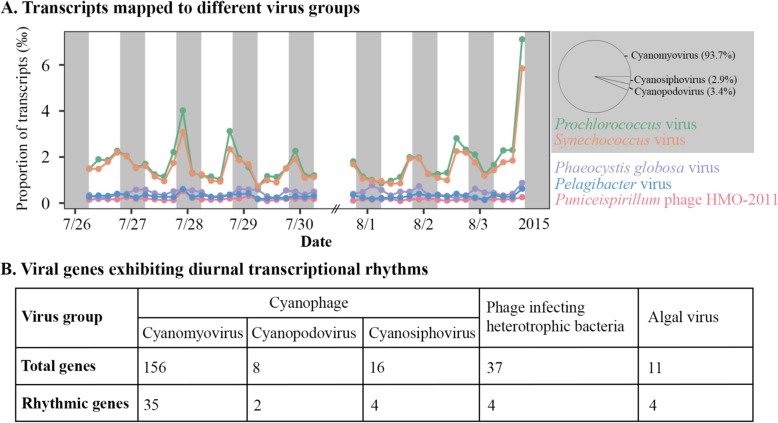
Background: Based on the peak expression times during infection, early, middle, and late genes have been characterized in viruses (cyanophages) that infect the unicellular cyanobacterium Prochlorococcus. Laboratory experiments show that some cyanophages can only replicate in the light and thus exhibit diurnal infection rhythms under light-dark cycles. Field evidence also suggests synchronized infection of Prochlorococcus by cyanophages in the oceans, which should result in progressive expression of cyanophage early, middle, and late genes. However, distinct temporal expression patterns have not been observed in cyanophage field populations.
Results: In this study, we reanalyzed a previous metatranscriptomic dataset collected in the North Pacific Subtropical Gyre. In this dataset, it was previously shown that aggregate transcripts from cyanophage scaffolds display diurnal transcriptional rhythms with transcript abundances decreasing at night. By mapping metatranscriptomic reads to individual viral genes, we identified periodically expressed genes from putative viruses infecting the cyanobacteria Prochlorococcus and Synechococcus, heterotrophic bacteria, and algae. Of the 41 cyanophage genes, 35 were from cyanomyoviruses. We grouped the periodically expressed cyanomyovirus genes into early, middle, and late genes based on the conserved temporal expression patterns of their orthologs in cyanomyovirus laboratory cultures. We found that the peak expression times of late genes in cyanophage field populations were significantly later than those of early and middle genes, which were similar to the temporal expression patterns of synchronized cyanophage laboratory cultures.
Conclusions: The significantly later peak expression times of late genes in cyanomyovirus field populations suggest that cyanophage infection of Prochlorococcus is synchronized in the North Pacific Subtropical Gyre. The night-time peak expression of late genes also suggests synchronized lysis of Prochlorococcus at night, which might result in synchronized release of dissolved organic matter to the marine food web. Video abstract.
Keywords: Cyanobacterium; Cyanophage; Diurnal rhythm; Light-dark cycle; Metatranscriptomics.
Conflict of interest statement
The authors declare that they have no competing interests.
Figures


References
-
- Benjamini Y, Hochberg Y. Controlling the false discovery rate: a practical and powerful approach to multiple testing. Journal of the Royal Statistical Society. Series B (Methodological) 1995;57:289–300.
-
- Breitbart M, Thompson LR, Suttle CA, Sullivan MB. Exploring the vast diversity of marine viruses. Oceanography. 2007;20:135–139.
-
- Clokie MR, Shan J, Bailey S, Jia Y, Krisch HM, West S, Mann NH. Transcription of a ‘photosynthetic’ T4-type phage during infection of a marine cyanobacterium. Environ Microbiol. 2006;8:827–835. - PubMed
Publication types
MeSH terms
Grants and funding
LinkOut - more resources
Full Text Sources
