. 2020 Jun 24;13(6):dmm042549.
doi: 10.1242/dmm.042549.

Zebrafish models of skeletal dysplasia induced by cholesterol biosynthesis deficiency

Affiliations

Zebrafish models of skeletal dysplasia induced by cholesterol biosynthesis deficiency

Rebecca A Anderson et al. Dis Model Mech. .

Abstract

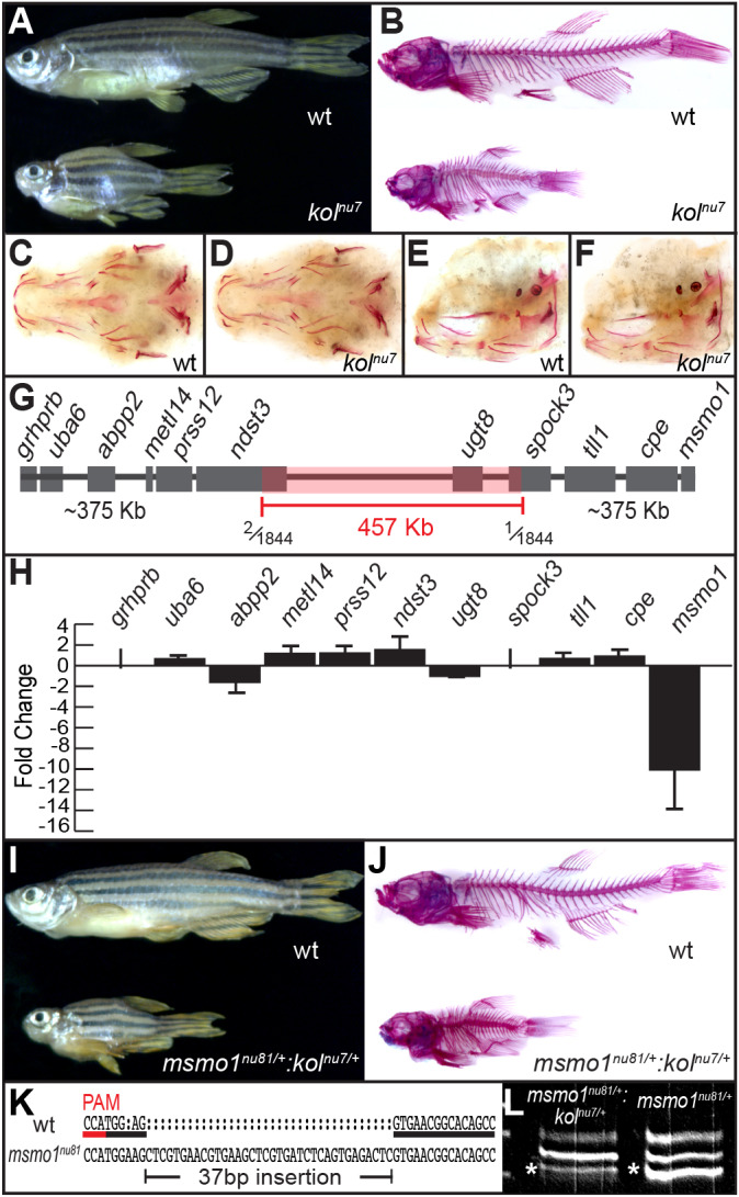
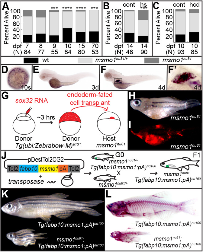
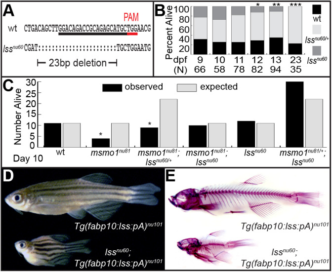
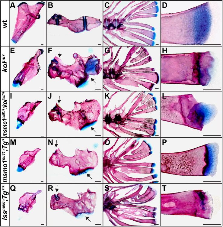
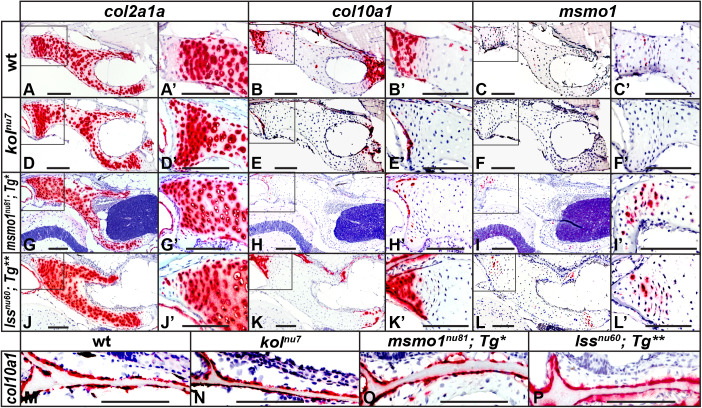
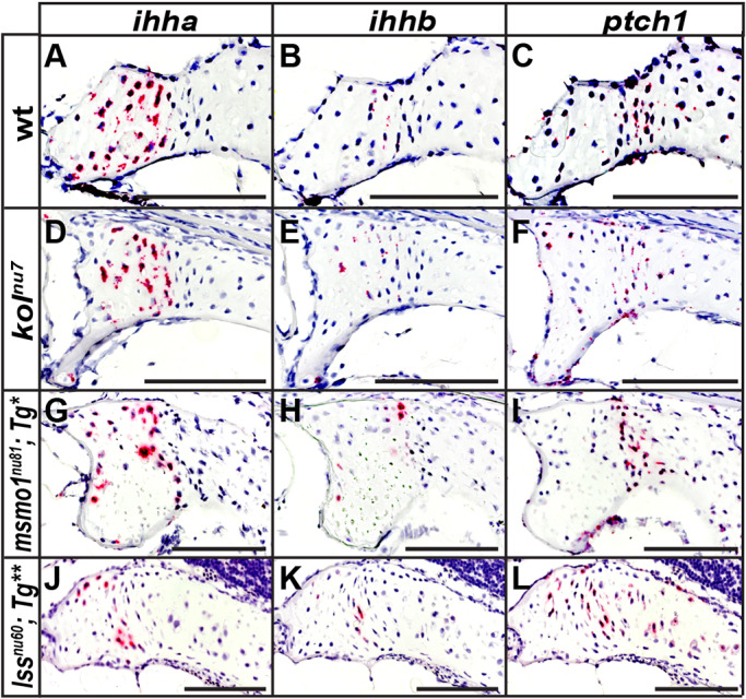
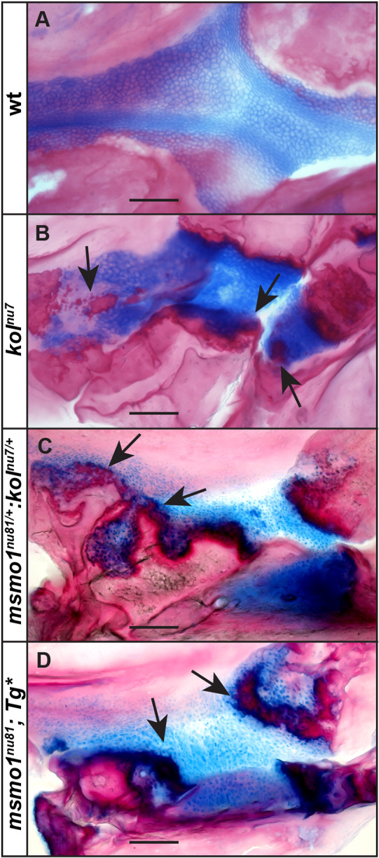
Human disorders of the post-squalene cholesterol biosynthesis pathway frequently result in skeletal abnormalities, yet our understanding of the mechanisms involved is limited. In a forward-genetic approach, we have found that a late-onset skeletal mutant, named kolibernu7 , is the result of a cis-acting regulatory mutation leading to loss of methylsterol monooxygenase 1 (msmo1) expression within pre-hypertrophic chondrocytes. Generated msmo1nu81 knockdown mutation resulted in lethality at larval stage. We demonstrated that this is a result of both cholesterol deprivation and sterol intermediate accumulation by creating a mutation eliminating activity of Lanosterol synthase (Lss). Our results indicate that double lssnu60;msmo1nu81 and single lssnu60 mutants survive significantly longer than msmo1nu81 homozygotes. Liver-specific restoration of either Msmo1 or Lss in corresponding mutant backgrounds suppresses larval lethality. Rescued mutants develop dramatic skeletal abnormalities, with a loss of Msmo1 activity resulting in a more-severe patterning defect of a near-complete loss of hypertrophic chondrocytes marked by col10a1a expression. Our analysis suggests that hypertrophic chondrocytes depend on endogenous cholesterol synthesis, and blocking C4 demethylation exacerbates the cholesterol deficiency phenotype. Our findings offer new insight into the genetic control of bone development and provide new zebrafish models for human disorders of the cholesterol biosynthesis pathway.

Keywords: Cholesterol; Chondrodysplasia punctata; Lss; Msmo1; Skeletal dysplasia; Zebrafish.


Conflict of interest statement

Competing interestsThe authors declare no competing or financial interests.

Figures

Fig. 1.
Fig. 1.
Late-onset skeletal defects observed in the kolibernu7 (kolnu7) mutant are the result of downregulation of msmo1 expression. (A) Compared to wild-type (wt) siblings (top), adult kolnu7 mutants display a reduced body length and small head size. wt n=300, kolnu7 n=300. (B) Whole-mount skeletal preparations reveal gross malformations and hyperossification throughout the adult kolnu7 craniofacial and axial skeleton after Alcian Blue (cartilage) and Alizarin Red (ossified bone and mineralized tissues) staining. wt n=100, kolnu7 n=100. (C-F) Early larval mutants do not display patterning defects or premature ossification. Whole-mount Alizarin Red staining of 4.7 mm (∼8 dpf) wt (C,E) and kolnu7. (D,F). Ventral view (C,D) and lateral view (E,F). wt n=3, kolnu7 n=4. (G) Positional cloning reveals that the kolnu7 locus is located to the ∼457 kb critical region flanked by polymorphic markers with one or two recombinants out of 1844 meioses, corresponding to a genetic distance of 0.16 cM. (H) Screen of gene expression using quantitative RT-PCR from RNA extracted from hypural complex of ∼18 mm SL kolnu7 and wt siblings. wt n=2, kolnu7 =2. Only msmo1 level was significantly different out of 11 tested genes, located in the ∼1.2 Mb region encompassing the kolnu7 locus. Initial screen results: grhprb not detected (ND); uba6 mean difference 0.64, s.d. 0.36; abpp2 −1.62, s.d. 1.00; mettl14 1.16, s.d. 0.68; prss12 1.20, s.d. 0.72; ndst3 1.51, s.d. 1.50; ugt8 −1.03, s.d. 0.07; spock3 ND; tll1 0.67, s.d. 0.27; cpe 0.90, s.d. 0.42; msmo1 −10.07, s.d. 4.32. Confirmation test of msmo1 expression (msmo1 −11.47, s.d. 8.49; P=0.0012), wt n=3, kolnu7 =5. Three technical replicates were included for all assays. Gene expression was normalized to the reference gene eefla1. Fold change was calculated using Livak method (Livak and Schmittgen, 2001). P-value calculated using unpaired Student's t-test on dCt values. (I) The msmo1nu81 mutant allele is not able to complement the kolnu7 mutation. Adult kolnu7/+:msmolnu81/+ transheterozygotes phenocopy the kolnu7 mutant. wt n=100, kolnu7/+:msmolnu81/+ n=100. (J) Whole-mount skeletal preparations reveal gross malformations throughout the kolnu7/+:msmolnu81/+ craniofacial and axial skeleton, similar to those observed in kolnu7. wt n=10, kolnu7/+:msmo1nu81/+ n=10. (K) The msmo1nu81 allele is the result of a 37 bp insertion, allowing for allele-specific expression analysis between msmo1nu81 and kolnu7. PAM, protospacer adjacent motif (underlined in red). (L) Strong downregulation of the kolnu7-linked allele (asterisks) in kolnu7/+:msmo1nu81/+ compared to the wt allele in msmo1nu81/+ suggests that the kolnu7 mutation is cis-acting. kolnu7/+:msmo1nu81/+ n=3, msmo1nu81/+ n=3. The top band is a heterodimer of wt and mutant strands.
Fig. 2.
Fig. 2.
The suppression of early lethality of the msmo1nu81 mutation yields adult fish with kolnu7-like phenotype. (A) Survival of larvae from msmo1nu81/+ in-crosses, from 7 dpf to 70 dpf. Most msmo1nu81 mutants die by 9 dpf. [7 dpf wild type (wt; black) n=19, heterozygotes (het; pale gray) n=49, knockout mutants (KO; dark gray) n=16, P=0.02794; 8 dpf wt n=21, het n=39, KO n=17, P=0.8065; 9 dpf wt n=12, het n=40, KO n=3, ***P=0.0008; 10 dpf wt n=37, het n=44, KO n=3, ****P<0.0001; 15 dpf wt n=26, het n=51, KO n=3, ****P<0.0001; 70 dpf wt n=18, het n=35, KO n=0, ***P=0.001.] (B) Overexpression of msmo1 driven by daily heat shock of the transgenic line Tg(hsp70l:msmo1:IRESnlsGFP)nu99 rescued the lethality of msmo1nu8 mutants. Transgenic screening based on cardiac GFP. (Control non-transgenic siblings 14 dpf wt n=14, het n=32, KO n=2, ***P=0.0035; transgenic siblings 14 dpf wt n=18, het n=44, KO n=28, P=0.3214.) cont, control; hs, heat shock. (C) Dietary cholesterol supplementation does not improve the survivability of msmo1nu81 mutants. Clutches from msmo1nu81/+ in-crosses were fed either a high-cholesterol diet (hcd) or a control standardized diet (cont) beginning at 5 dpf until collection at 10 dpf. (HCD wt n=26, het n=50, KO n=9, P=0.0089; control diet wt n=22, het n=66, KO n=5, P<0.0001.) All two-tailed P-values were calculated using chi-squared test. (D-F′) Whole-mount in situ hybridization during the first 5 days of development shows msmo1 expression predominately in the yolk syncytial layer (YSL) and liver. Expression is first detected during early somitogenesis in the YSL (D) and continues there at 3 dpf (E). At 4 dpf, strong expression is observed in the differentiated liver (F,F′). (G-I) Generation of msmo1nu81/wild-type chimeras using endoderm replacement rescues early lethality of msmo1nu81 mutants and reveals a strong kolnu7-like phenotype. (G) Schematic of the procedure. Wild-type Tg(ubi:Zebrabow-M)a131 donor embryos were injected with sox32 RNA to force an endodermal fate. At high stage (∼3 hpf), cells were transplanted from donor to host embryos collected from msmo1nu81/+ in-crosses. (H) Surviving msmo1nu81 mutants display a strong kolnu7-like phenotype. msmo1nu81 n=4. (I) The majority of organs of endodermal origin displayed a high enrichment in transplanted Tg(ubi:Zebrabow-M)a131 cells (red). msmo1nu81 n=4. (J-L) Liver-specific msmo1 expression in msmo1nu81 mutants rescues early lethality and produces juvenile msmo1nu81 mutants with strong kolnu7-like phenotype. (J) Liver-specific regulatory element fabp10a was used to drive msmo1 expression in msmo1nu81 mutants. (K) Adult Tg(fabp10a:msmo1:pA)nu100;msmo1nu81 mutants phenocopy kolnu7 mutant. Tg(fabp10:msmo1:pA)nu100 n=50, Tg(fabp10a:msmo1:pA)nu100;msmo1nu81 n=50. (L) Whole-mount skeletal preparations reveal gross malformations throughout Tg(fabp10a:msmo1:pA)nu100;msmo1nu81 craniofacial and axial skeleton, similar to those observed in kolnu7. Tg(fabp10:msmo1:pA)nu100 n=3, Tg(fabp10a:msmo1:pA)nu100;msmo1nu81 n=7.
Fig. 3.
Fig. 3.
Loss of Lss activity phenotype is epistatic to msmo1nu81 mutation. (A) The lssnu60 allele. PAM, protospacer adjacent motif (underlined in red). (B) Survival analysis of clutches of lssnu60/+ in-crosses, from 9 dpf to 23 dpf, reveals that most lssnu60 mutants die by 12 dpf. [9 dpf wild type (wt; black) n=21, heterozygotes (het; pale gray) n=30, knockout mutants (KO; dark gray) n=15, P=0.4412; 10 dpf wt n=15, het n=27, KO n=16, P=0.8563; 11 dpf wt n=20, het n=44, KO n=14, P=0.3320; 12 dpf wt n=24, het n=48, KO n=10, *P=0.0277; 13 dpf wt n=33, het n=51, KO n=10, **P=0.0026; 23 dpf wt n=8, het n=27, KO n=0, ***P=0.0009.] (C) Survival analysis of clutches of msmo1nu81/+;lssnu60/+ in-crosses reveals that loss of Lss activity increases survivability of msmo1nu81 mutants at 10 dpf. [wt observed (O) n=11, expected (E) n=11; msmo1nu81 O n=4, E n=11; msmo1nu81;lssnu60/+ O n=9, E n=22; msmo1nu81;lssnu60 O n=10, E n=11; lssnu60 O n=12, E n=11; msmo1nu81/+;lssnu60 O n=30, E n=22; msmo1nu81/+;lssnu60/+ O n=40, E n=44; msmo1nu81/+ O n=18, E n=22; lssnu60/+ O n=18, E n=22; *P=0.0431.] Two-tailed P-values were calculated using chi-squared test. (D,E) Suppression of early lethality of lssnu60 mutants by liver-specific expression of lss. (D) Adult Tg(fabp10:lss:pA)nu101;lssnu60 mutants phenocopy kolnu7. Tg(fabp10:lss:pA)nu101 n=12, Tg(fabp10:lss:pA)nu101;lssnu60 n=12. (E) Whole-mount skeletal preparations reveal gross malformations throughout Tg(fabp10:lss:pA)nu101;lssnu60 craniofacial and axial skeleton, similar to those observed in kolnu7. Tg(fabp10:lss:pA)nu101 n=3, Tg(fabp10:lss:pA)nu101;lssnu60 n=5.
Fig. 4.
Fig. 4.
Loss of Msmo1 and Lss activity is associated with abnormal endochondral bone formation. (A-T) The hyomandibular (A,E,I,M,Q), ceratohyal (B,F,J,N,R) and hypural complex (C,D,G,H,K,L,O,P,S,T) were compared between wild type (A-D), kolnu7 (E-H), kolnu7/+:msmolnu81/+ (I-L), Tg(fabp10a:msmo1:pA)nu100;msmo1nu81 (M-P) and Tg(fabp10:lss:pA)nu101;lssnu60 (Q-T). (E-T) The endochondral bones of the mutants are compressed and irregularly shaped with ectopic ossification and fusions throughout the growth plates, marked with arrows. Images represent disarticulated whole-mount adult skeletal preparations at ∼4 months of age. Scale bars: 100μm. Tg*, Tg(fabp10:msmo1:pA)nu100; Tg**, Tg(fabp10:lss:pA)nu101. wt n=4, kolnu7 n=4, kolnu7/+:msmolnu81/+ n=4, Tg(fabp10a:msmo1:pA)nu100;msmo1nu81 n=4, Tg(fabp10:lss:pA)nu101;lssnu60 n=4.
Fig. 5.
Fig. 5.
Loss of Msmo1 and Lss activities disrupts growth plate patterning. (A-P) Expression domains of col2a1a (A,D,G,J), col10a1a (B,E,H,K,M-P) and msmo1 (C,F,I,L) were analyzed within growth plates of wild type (A-C), kolnu7 (D-F), Tg(fabp10a:msmo1:pA)nu100;msmo1nu81 (G-I) and Tg(fabp10:lss:pA)nu101;lssnu60 (J-L) using RNAscope in situ hybridization. (A) Strong col2a1a expression is observed in round and stacked chondrocytes in wild type, corresponding to presumed resting and proliferating chondrocytes. (B) Expression of col10a1a in wild type is observed complementary to col2a1a expression in presumed hypertrophic chondrocytes. The expression of col2a1a is expanded in kolnu7 (D) and Tg(fabp10a:msmo1:pA)nu100;msmo1nu81 (G) growth plates, with a near-complete loss of col10a1a expression in kolnu7 (E) and Tg(fabp10a:msmo1:pA)nu100;msmo1nu81 (H). Expression of col2a1a (J) and col10a1a (K) in Tg(fabp10:lss:pA)nu101;lssnu60 appears less affected when compared to kolnu7(D,E) and Tg(fabp10a:msmo1:pA)nu100;msmo1nu81 (G,H). Expression of msmo1 is detected primarily in pre-hypertrophic chondrocytes of wild type (C); msmo1 expression is undetectable in kolnu7 growth plates (F), but is upregulated in transgenically rescued msmo1nu81 (I) and lssnu60 (L) mutants. (M-P) Expression of col10a1a in osteoblasts is unchanged between wild type and mutants. Images represent paraffin sections of the pterotic at 2 months of age (∼15 mm SL). Boxed areas in A-L are shown at higher magnification in A′-L′. Scale bars: 100 μm. Tg*, Tg(fabp10:msmo1:pA)nu100; Tg**, Tg(fabp10:lss:pA)nu101. wt n=6, kolnu7 n=6, Tg(fabp10a:msmo1:pA)nu100;msmo1nu81 n=3, Tg(fabp10:lss:pA)nu101;lssnu60 n=3.
Fig. 6.
Fig. 6.
Ihh signaling within endochondral growth plates is present despite loss of Msmo1 and Lss activity. (A-L) Expression domains of ihha (A,D,G,J), ihhb (B,E,H,K) and ptch1 (C,F,I,L) were analyzed within growth plates of wild type (A-C), kolnu7 (D-F), Tg(fabp10a:msmo1:pA)nu100;msmo1nu81 (G-I) and Tg(fabp10:lss:pA)nu101;lssnu60 (J-L) using RNAscope in situ hybridization. (A-C) In wild type, expression of ihha and ihhb is observed in the pre-hypertrophic chondrocyte zone, with ptch1 expression seen to either side of these domains. (D-L) Expression of ihha, ihhb and ptch1 is present within mutant growth plates and similar to domains observed in wild type. Images represent paraffin sections of the pterotic at 2 months of age (∼15 mm SL). Scale bars: 100 μm. Tg*, Tg(fabp10:msmo1:pA)nu100; Tg**, Tg(fabp10:lss:pA)nu101. wt n=3, kolnu7 n=3, kolnu7/+:msmolnu81/+ n=3, Tg(fabp10a:msmo1:pA)nu100;msmo1nu81 n=3, Tg(fabp10:lss:pA)nu101;lssnu60 n=3.
Fig. 7.
Fig. 7.
New zebrafish models for CDP. (A-D) Abnormal ossification, marked with arrows, within endochondral growth plates of kolnu7 (B), kolnu7/+:msmolnu81/+ (C) and Tg(fabp10a:msmo1:pA)nu100;msmo1nu81 (D) mutant phenotypes resembles defects observed in patients with chondrodysplasia punctata. Images represent lateral views of palatoquadrate growth plates of craniofacial bones from disarticulated whole-mount adult skeletal preparations at ∼4 months of age. Scale bars: 100 μm. Tg*, Tg(fabp10:msmo1:pA)nu100. wt n=4, kolnu7 n=6, kolnu7/+:msmolnu81/+ n=4, Tg(fabp10a:msmo1:pA)nu100;msmo1nu81 n=4.

References

    1. Ačimovič J., Goyal S., Košir R., Goličnik M., Perše M., Belič A., Urlep Z., Guengerich F. P. and Rozman D. (2016). Cytochrome P450 metabolism of the post-lanosterol intermediates explains enigmas of cholesterol synthesis. Sci. Rep. 6, 28462 10.1038/srep28462 - DOI - PMC - PubMed
    1. Albuisson J., Isidor B., Giraud M., Pichon O., Marsaud T., David A., Le Caignec C. and Bezieau S. (2011). Identification of two novel mutations in Shh long-range regulator associated with familial pre-axial polydactyly. Clin. Genet. 79, 371-377. 10.1111/j.1399-0004.2010.01465.x - DOI - PubMed
    1. Avaron F., Hoffman L., Guay D. and Akimenko M. A. (2006). Characterization of two new zebrafish members of the hedgehog family: atypical expression of a zebrafish indian hedgehog gene in skeletal elements of both endochondral and dermal origins. Dev. Dyn. 235, 478-489. 10.1002/dvdy.20619 - DOI - PubMed
    1. Besnard T., Sloboda N., Goldenberg A., Kury S., Cogne B., Breheret F., Trochu E., Conrad S., Vincent M., Deb W. et al. (2019). Biallelic pathogenic variants in the lanosterol synthase gene LSS involved in the cholesterol biosynthesis cause alopecia with intellectual disability, a rare recessive neuroectodermal syndrome. Genet. Med. 21, 2025 10.1038/s41436-019-0445-x - DOI - PubMed
    1. Bird N. C. and Mabee P. M. (2003). Developmental morphology of the axial skeleton of the zebrafish, Danio rerio (Ostariophysi: Cyprinidae). Dev. Dyn. 228, 337-357. 10.1002/dvdy.10387 - DOI - PubMed

Publication types

MeSH terms