. 2020 May 19;11(3):e00937-20.
doi: 10.1128/mBio.00937-20.

A New Pneumococcal Capsule Type, 10D, is the 100th Serotype and Has a Large cps Fragment from an Oral Streptococcus

Affiliations

A New Pneumococcal Capsule Type, 10D, is the 100th Serotype and Has a Large cps Fragment from an Oral Streptococcus

Feroze Ganaie et al. mBio. .

Abstract

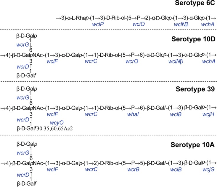
Streptococcus pneumoniae (pneumococcus) is a major human pathogen producing structurally diverse capsular polysaccharides. Widespread use of highly successful pneumococcal conjugate vaccines (PCVs) targeting pneumococcal capsules has greatly reduced infections by the vaccine types but increased infections by nonvaccine serotypes. Herein, we report a new and the 100th capsule type, named serotype 10D, by determining its unique chemical structure and biosynthetic roles of all capsule synthesis locus (cps) genes. The name 10D reflects its serologic cross-reaction with serotype 10A and appearance of cross-opsonic antibodies in response to immunization with 10A polysaccharide in a 23-valent pneumococcal vaccine. Genetic analysis showed that 10D cps has three large regions syntenic to and highly homologous with cps loci from serotype 6C, serotype 39, and an oral streptococcus strain (S. mitis SK145). The 10D cps region syntenic to SK145 is about 6 kb and has a short gene fragment of wciNα at the 5' end. The presence of this nonfunctional wciNα fragment provides compelling evidence for a recent interspecies genetic transfer from oral streptococcus to pneumococcus. Since oral streptococci have a large repertoire of cps loci, widespread PCV usage could facilitate the appearance of novel serotypes through interspecies recombination.IMPORTANCE The polysaccharide capsule is essential for the pathogenicity of pneumococcus, which is responsible for millions of deaths worldwide each year. Currently available pneumococcal vaccines are designed to elicit antibodies to the capsule polysaccharides of the pneumococcal isolates commonly causing diseases, and the antibodies provide protection only against the pneumococcus expressing the vaccine-targeted capsules. Since pneumococci can produce different capsule polysaccharides and therefore reduce vaccine effectiveness, it is important to track the appearance of novel pneumococcal capsule types and how these new capsules are created. Herein, we describe a new and the 100th pneumococcal capsule type with unique chemical and serological properties. The capsule type was named 10D for its serologic similarity to 10A. Genetic studies provide strong evidence that pneumococcus created 10D capsule polysaccharide by capturing a large genetic fragment from an oral streptococcus. Such interspecies genetic exchanges could greatly increase diversity of pneumococcal capsules and complicate serotype shifts.

Keywords: Streptococcus pneumoniae; capsule; genetic exchange; vaccine.


Figures

FIG 1
FIG 1
Serological profile of strain Cam853 and other pneumococcal serotypes by flow cytometric analysis. Histograms show fluorescence of a bacterial strain (indicated at the top of each column of histograms) after staining with the indicated serologic reagents (indicated to the left of each row of histograms). Pneumococcal strains SSISP39, SSISP10A, SSISP10B, SSISP10C, SSISP10F, and MJC705 represents serotypes 39, 10A, 10B, 10C, 10F, and 6C, respectively. The solid black lines indicate the fluorescence obtained with primary and secondary antibodies. The gray shaded areas represent control binding (secondary antibody alone). The x axes show the log fluorescence intensity, and the y axes show the number of events (cell counts). Hyp10AG1 is the serotype 10A-specific monoclonal antibody (MAb). All other reagents were obtained from Statens Serum Institut (SSI) (FS10d, factor serum 10d). Each strain was tested three times, and a representative result was shown.
FIG 2
FIG 2
(A) Purification of serotype 10D capsular polysaccharide. Anion-exchange fractions (x axis) were tested for 10D capsule polysaccharide (PS) (solid diamonds), OD260 (open squares), and OD280 (triangles). 10D polysaccharide was determined by an inhibition ELISA. OD260 and OD280 reflect nucleic acid and protein contaminations, respectively. Fraction 15 (indicated by an arrow) was selected for nuclear magnetic resonance (NMR) studies. (B) One-dimensional 1H NMR spectra of a selected region of the 1H NMR spectra of 6C, 10A, 39. and 10D capsule polysaccharides. Purified capsular polysaccharides of serotypes 39, 10A, and 6C were procured from Statens Serum Institut. Asterisks indicate cell wall polysaccharide-specific peaks. The NMR spectrum of anomeric carbons (4.6 to 5.6 ppm) indicates that 10D capsule polysaccharide is chemically distinct from 39, 10A, and 6C. SGT, serogroup/type.
FIG 3
FIG 3
Two-dimensional nuclear magnetic resonance (NMR) spectra of serotype 10D polysaccharide. 1H-13C HMQC spectra of anomeric carbons and the other carbons are shown in panels A and B, respectively. (C) 1H-31P HMBC spectrum of the ribitol phosphate region. NMR spectra were obtained at 45°C. In the peak labels, the letter indicates the sugar residue (see Table 4), and the number denotes the carbon position on each unit. Peak assignments are shown in Table 4. The asterisk denotes an unassigned peak. Horizontal dashed lines connect the two proton signals associated with the sugar residue at the indicated carbon position.
FIG 4
FIG 4
Structure of the serotype 10D capsule polysaccharide in comparison to serotype 6C, 39, and 10A polysaccharide. The structure of the repeating unit of the polysaccharide from each serotype is shown along with the genes (shown in blue) responsible for biosynthesis of the chemical structure. Serotype 10D shares the two glucose residues and their corresponding glycosyltransferase genes with serotype 6C. Ribitol phosphotransferase wcrO10D is responsible for the ribitol (5→P→6) glucose linkage. The remaining structure on the left is similar to those in serotypes 39 and 10A, except that the wcyO gene mediates O-acetylation in serotype 39, and wcrC10A mediates (1→2) linkage in serotype 10A. The structural similarity in Galp, Galf, and GalNAc residues in serotypes 10D, 39, and 10A explains the serological cross-reactivity with FS10d and Hyp10AG1.
FIG 5
FIG 5
23-valent pneumococcal polysaccharide vaccine (PPV23) elicits opsonic antibodies to serotype 10D. (A) Capacity to opsonize serogroup/type (SGT) 10A (left) or 10D (right) with sera from six adults before and after PPV23 immunization. The dashed black lines represent the mean opsonophagocytic indices. Error bars indicate standard deviations. Data were analyzed by the Mann-Whitney test. (B) Capacity of PPV23 immune sera to opsonize SGT 10A and 10D in the presence of OBB buffer (left), 10A PS (middle). or unrelated PS (right). Inhibitor concentrations were 200 μg/ml in OBB. Values are means ± standard deviations (error bars). Data were analyzed by a one-way analysis of variance (ANOVA) with Tukey’s multiple-comparison test. Values that are significantly different are indicated by a bar and asterisks as follows: **, P < 0.005; ***, P < 0.001; ****, P < 0.0001). PS, polysaccharide.
FIG 6
FIG 6
Phylogenetic analysis of the wcrO10D gene. The phylogenetic tree shows that the wcrO10D gene has the highest homology (90% identical at the nucleotide level and 94% at the amino acid level) with the S. mitis SK145 gene RS00925. All the genes from pneumococcal and nonpneumococcal strains depicted in the tree belong to pfam04991 (https://www.ncbi.nlm.nih.gov/Structure/cdd/wrpsb.cgi). The question mark symbol indicates that the gene function is unknown. The length of the scale bar represents the estimated evolutionary divergence between species based on average genetic distance. A scale bar of 0.1 indicates 10% nucleotide substitution per site. Nucleotide and translated amino acid sequences below 45% and 30% identity, respectively, were eliminated from the analysis. RS06905, RS00925, RS08245, 9626_0115, 9962_0662, and RS05515 are locus tags. The NCBI accession numbers of all the strains used in phylogenetic analysis are provided in Table S1 in the supplemental material. SGT, serogroup/type.
FIG 7
FIG 7
Potential evolutionary origin of serotype 10D capsule biosynthetic loci (cps). (A) Comparison of 10D cps (SRA accession no. ERR051587) (21) with 6C cps (GenBank accession no. EF538714), SK145 cps (NZ_JYGS01000001.1; contig 1), and 39 cps (CR931711) using dot plot analysis. The x axis shows the 10D cps gene arrangement; 6C, SK145, and 39 cps loci are plotted on the y axis. For strain SK145, reverse complement sequence was used for comparison. The numbers on the x and y axes relate to arbitrary nucleotide base numbers (in kilobases) to show relative size. The solid black lines represent the homologous regions, with the percentage nucleotide identity indicated above the line. The vertical gray-shaded areas indicate overlap regions for the potential recombination events I and II. (B) The nucleotide sequences of cps loci for serotype 6C, serotype 10D, and strain SK145 at the transition region for the potential recombination event I. The sequence in bold type indicates the overlap region. Underlined nucleotides indicate the wciNβ6C and wciNβ10D translation stop codons. Lowercase letters in 10D cps indicate the nucleotide mismatches. The base numbers indicate the actual nucleotide position in the corresponding cps loci. The gray-shaded area indicates the region of homology “upstream” (cps 6C and cps 10D) and “downstream” (cps 10D and cps SK145) of the potential recombination site. The dashed horizontal lines indicate the genes involved in the potential recombination event I.

References

    1. Pasteur L. 1881. Note sur la maladie nouvelle provoquée par la salive d’un enfant mort de la rage. Bull Acad Méd (Paris) 10:94–103.
    1. GBD 2015 LRI Collaborators. 2017. Estimates of the global, regional, and national morbidity, mortality, and aetiologies of lower respiratory tract infections in 195 countries: a systematic analysis for the Global Burden of Disease Study 2015. Lancet Infect Dis 17:1133–1161. doi:10.1016/S1473-3099(17)30396-1. - DOI - PMC - PubMed
    1. Avery OT, Dubos R. 1931. The protective action of a specific enzyme against type III pneumococcus infection in mice. J Exp Med 54:73–89. doi:10.1084/jem.54.1.73. - DOI - PMC - PubMed
    1. Avery OT, Macleod CM, McCarty M. 1944. Studies on the chemical nature of the substance inducing transformation of pneumococcal types: induction of transformation by a desoxyribonucleic acid fraction isolated from pneumococcus type III. J Exp Med 79:137–158. doi:10.1084/jem.79.2.137. - DOI - PMC - PubMed
    1. Andrews NJ, Waight PA, Burbidge P, Pearce E, Roalfe L, Zancolli M, Slack M, Ladhani SN, Miller E, Goldblatt D. 2014. Serotype-specific effectiveness and correlates of protection for the 13-valent pneumococcal conjugate vaccine: a postlicensure indirect cohort study. Lancet Infect Dis 14:839–846. doi:10.1016/S1473-3099(14)70822-9. - DOI - PubMed

Publication types

LinkOut - more resources