Review
. 2020 May 19;11(1):70.
doi: 10.1186/s13244-020-00877-4.

Evaluation of the bronchial arteries: normal findings, hypertrophy and embolization in patients with hemoptysis

Affiliations
Review

Evaluation of the bronchial arteries: normal findings, hypertrophy and embolization in patients with hemoptysis

João Almeida et al. Insights Imaging. .

Abstract

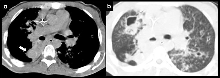
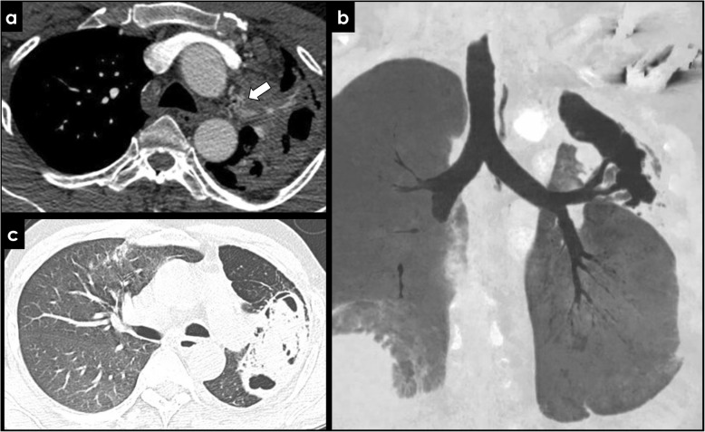
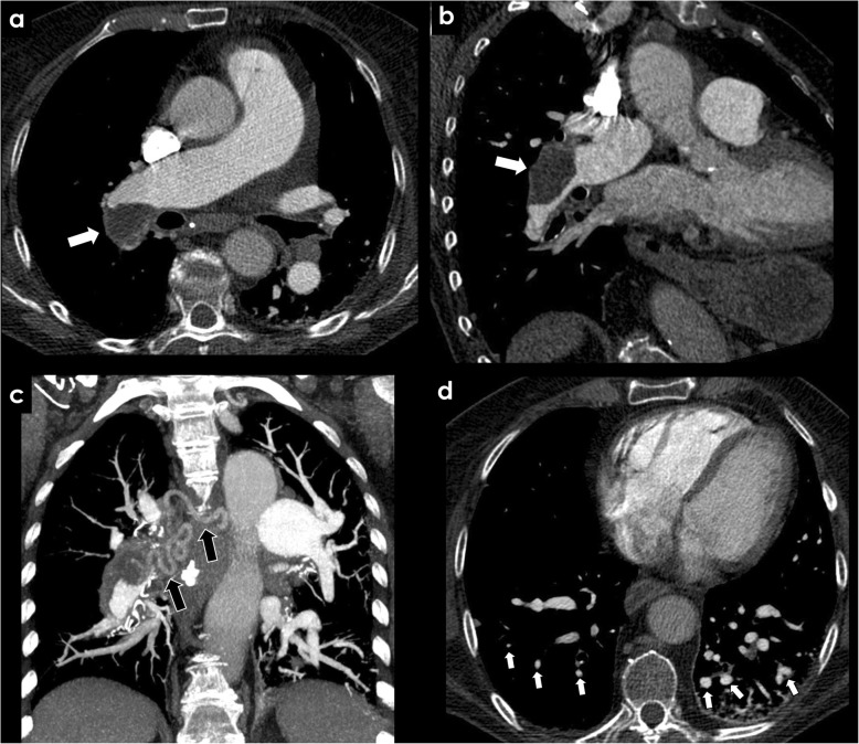
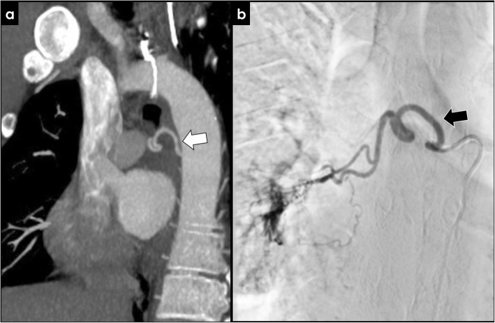
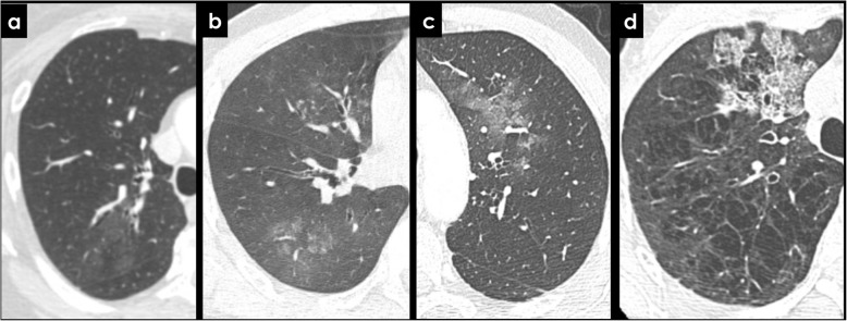
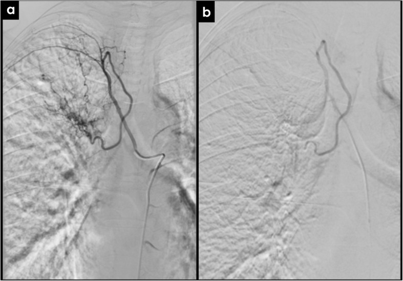
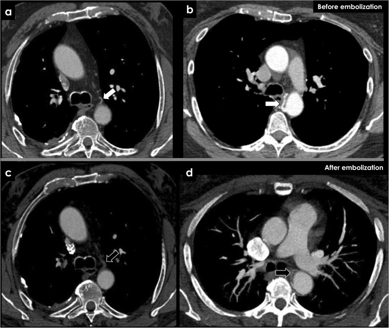
The enlargement of the bronchial arteries occurs in a multitude of congenital and acquired diseases and is responsible for the majority of cases of hemoptysis. In this review, we provide a simplified imaging approach to the evaluation of the bronchial arteries. We highlight the anatomy and function of the bronchial arteries, typical imaging findings, how to recognize bronchial artery dilatation, and its underlying causes. Contrast-enhanced computer tomography plays a major role in diagnosing bronchial artery enlargement and also improves treatment planning. Bronchial artery embolization has proven to be effective in controlling the potential hazardous hemoptysis.

Keywords: Angiography; Bronchial arteries; Computer tomography; Embolization; Hemoptysis.


Conflict of interest statement

The authors declare that they have no competing interests

Figures

Fig. 1
Fig. 1
Origin of orthotopic bronchial arteries. Axial (a) and sagital (b) CECT view of an orthotopic bronchial artery (black arrows) originating from the descending thoracic aorta (DA) at the T5–T6 vertebral plane, near the carinal level
Fig. 2
Fig. 2
Variations of the bronchial arteries. Bronchial arteries may have a great variability in terms of number. The most common patterns are two left and one right bronchial artery (type 1, most common), one artery in each side (type 2), two right and two left bronchial arteries (type 3), and two right and one left bronchial artery (type 4). Usually, the right bronchial artery originates from an intercostal-bronchial trunk (ICBT). RBA, right bronchial artery; LBA, left bronchial artery; ICBT, intercostal-bronchial trunk; ICA, intercostal artery
Fig. 3
Fig. 3
Imaging of the bronchial arteries. CECT findings of normal bronchial arteries (a, arrow), with a reduced caliber inferior to 1,5 mm. The remaining images show a patient with hypertrophy of the bronchial arteries, with a caliber exceeding 2 mm (b, c, d; arrows). MIP images (b, d) are useful when depicting small vessels like the bronchial arteries, displaying its tortuous course in a single image. The patient had a long-term history of bronchiectasis
Fig. 4
Fig. 4
Congenital causes of BAH—Tetralogy of Fallot and pulmonary agenesis. Significant bronchial artery hypertrophy (white arrows) in a patient with tetralogy of Fallot (ac) and in a patient with pulmonary artery agenesis (d)—“image b” shows a ventricular septal defect, overriding aorta and right ventricular hypertrophy, features of tetralogy of Fallot. The black arrow in “image d” depicts the absence of the pulmonary artery. RV, right ventricle; LV, left ventricle; AA, ascending aorta
Fig. 5
Fig. 5
Congenital causes of BAH—ALCAPA syndrome. Patient with an ALCAPA syndrome. CECT showing an anomalous left coronary artery arising from the pulmonary artery (a, black arrow) with associated bronchial artery dilatation (white arrows). PA, pulmonary artery; DA, descending aorta
Fig. 6
Fig. 6
Acquired causes of BAH—bronchiectasis. Patient with a massive left bronchial artery dilatation (a, white arrows) reaching 16 mm of caliber (b), related to the long-term history of bronchiectasis in the left lower lobe (c)
Fig. 7
Fig. 7
Acquired causes of BAH—cystic fibrosis. Ectopic dilated bronchial artery arising from the distal portion of the aortic arch (a, b; arrows) in a patient with cystic fibrosis. Typical pulmonary manifestations of cystic fibrosis are well depicted in the image (c)
Fig. 8
Fig. 8
Rasmussen’s aneurysm. Active TB can cause hemoptysis due to rupture of a Rasmussen’s aneurysm (a, white arrow), which is a pseudo-aneurysm of the pulmonary artery adjacent to a cavity in patients with pulmonary TB (b)
Fig. 9
Fig. 9
Acquired causes of BAH—tuberculosis and aspergilloma. Left bronchial artery enlargement (a, white arrow) in a patient with an aspergilloma complicating a left upper lobe cavity from tuberculosis (c). Image b represents the same patient 4 years before demonstrating an aerated cavity without signs of fungal colonization
Fig. 10
Fig. 10
Acquired causes of BAH—chronic thromboembolic disease. CECT findings in a patient with history of chronic thromboembolic disease. A peripheral filling defect with obtuse margins is seen in the right pulmonary artery (a, b; white arrows), with an asymmetric caliber of distal vessels (d, white arrows) and a significant right bronchial artery hypertrophy (c, black arrows)
Fig. 11
Fig. 11
Bronchial artery hypertrophy. Imaging findings of bronchial artery hypertrophy with CECT (a, white arrow) and angiography (b, black arrow). Hypervascularity and bronchial artery-pulmonary artery shunt are usually well illustrated with angiography (b)
Fig. 12
Fig. 12
CT findings of pulmonary hemorrhage. Hazy ground-glass infiltrates (a, b, c) representing intra-alveolar hemorrhage in patients with hemoptysis. Also note endobronchial filling in b and crazy paving pattern in d
Fig. 13
Fig. 13
Bronchial artery embolization. Selective catheter angiography before (a) and after (b) successful right bronchial artery embolization in a patient with hemoptysis in the context of chronic thromboembolic disease
Fig. 14
Fig. 14
CECT findings of bronchial arteries before and after embolization. After an effective bronchial artery embolization, absence of enhancement (c, black arrow) and caliber reduction (d, black arrow) are usual CECT findings. We can compare it to the imaging findings before the embolization (a, b; white arrows)

References

    1. Bruzzi John F., Rémy-Jardin Martine, Delhaye Damien, Teisseire Antoine, Khalil Chadi, Rémy Jacques. Multi–Detector Row CT of Hemoptysis. RadioGraphics. 2006;26(1):3–22. - PubMed
    1. Pump KK. Distribution of bronchial arteries in the human lung. Chest. 1972;62:447–451. - PubMed
    1. Jean-Baptiste E. Clinical assessment and management of massive hemoptysis. Crit Care Med. 2000;28:1642–1647. - PubMed
    1. Tanomkiat W, Tanisaro K. Radiographic relationship of the origin of the bronchial arteries to the left main bronchus. J Thorac Imaging. 2003;18:27–33. - PubMed
    1. Hartmann Ieneke J. C., Remy-Jardin Martine, Menchini Laura, Teisseire Antoine, Khalil Chadi, Remy Jacques. Ectopic origin of bronchial arteries: assessment with multidetector helical CT angiography. European Radiology. 2007;17(8):1943–1953. - PubMed

LinkOut - more resources