Review
. 2020 Oct;38(10):922-933.
doi: 10.1007/s11604-020-00990-w. Epub 2020 May 19.

Clinical applications of digital angiography with the harmonization function in body interventional radiology

Affiliations
Review

Clinical applications of digital angiography with the harmonization function in body interventional radiology

Hidekatsu Tateishi et al. Jpn J Radiol. 2020 Oct.

Abstract

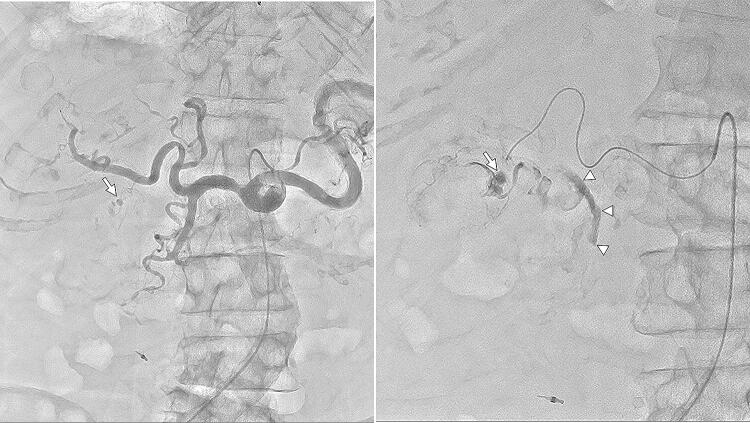
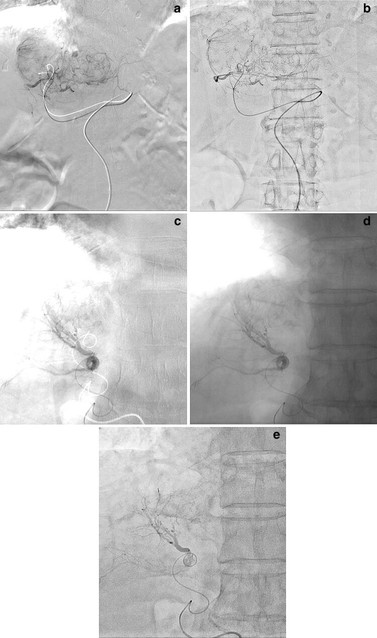
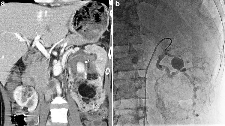
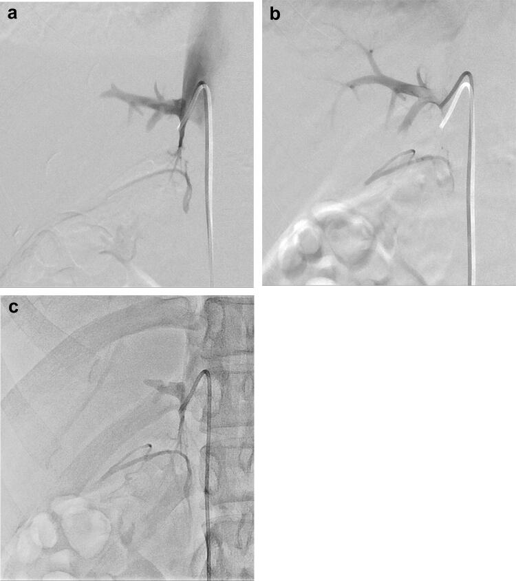
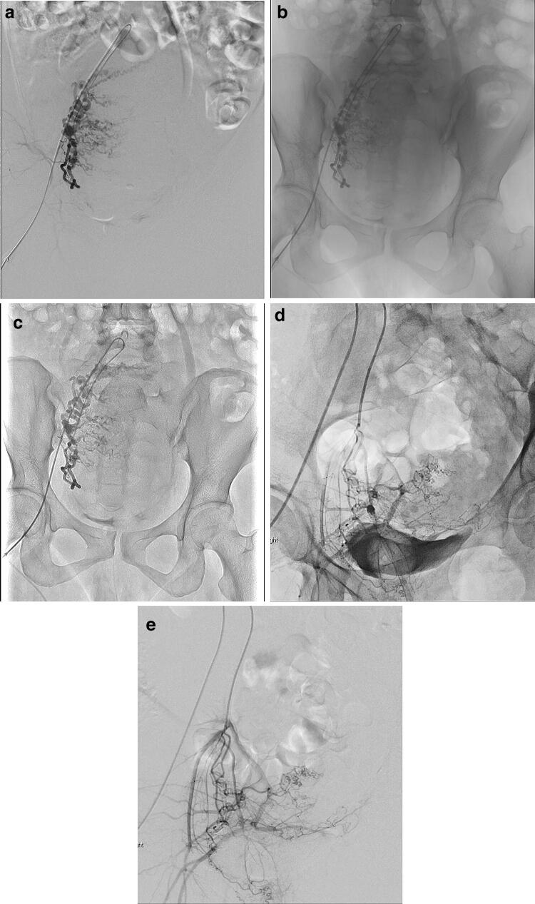
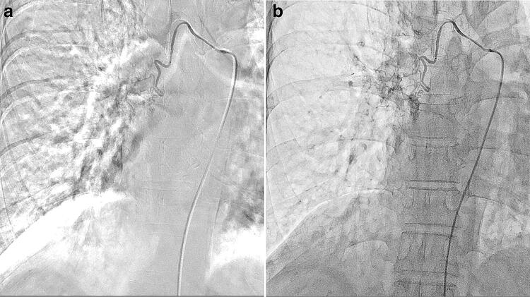
Digital subtraction angiography (DSA) is frequently applied in interventional radiology (IR). When DSA is not useful due to misregistration, digital angiography (DA) as an alternative option is used. In DA, the harmonization function (HF) works in real time by harmonizing the distribution of gray steps or reducing the dynamic range; thus, it can compress image gradations, decrease image contrast, and suppress halation artifacts. DA with HF as a good alternative to DSA is clinically advantageous in body IR for generating DSA-like images and simultaneously reducing various motion artifacts and misregistrations caused by patient body motion, poor breath-holding, bowel and ureter peristalsis, and cardiac pulsation as well as halation artifacts often stemming from the lung field. Free-breath DA with HF can improve body IR workflow and decrease the procedure time by reducing the risk of catheter dislocation and using background structures as anatomical landmarks, demonstrating reduced radiation exposure relative to DSA. Thus, HF should be more widely and effectively utilized for appropriate purposes in body IR. This article illustrates the basic facts and principles of HF in DA, and demonstrates clinical advantages and limitations of this function in body IR.

Keywords: Digital angiography; Dynamic density optimization; Harmonization function; Interventional radiology; Radiation exposure.


Figures

Fig. 1
Fig. 1
Stepping DA of the lower extremity with HF in an 84-year-old male with peripheral arterial disease. Stepping DA with HF following a single injection of contrast media well delineates the arteries of the whole lower extremity with less of an overlapped background relative to without HF
Fig. 2
Fig. 2
Rotational angiography of the right hand in a 43-year-old female with arteriovenous malformation of the right palm. While difficult to complete using DSA (a), DA with HF (b; Supplementary movie 1) facilitated the easy acquisition of rotational angiography to reveal a detailed three-dimensional relationship of such anatomically complicated vascular pathologies, particularly with reference to the carpal bones
Fig. 3
Fig. 3
IVC filter placement in a 22-year-old male with deep venous thrombosis. DSA of the IVC (a) well delineates the confluence sites of the renal veins (arrows) without visualization of vertebrae. In contrast, DA (b, c) visualizes vertebrae as anatomical landmarks to determine the release site of the filter. Compared with DA without HF (b), DA with HF (c) better delineates these confluence sites with less of an overlapped background
Fig. 4
Fig. 4
TAE for gastrointestinal hemorrhage in a 57-year-old male with hemorrhage from ascending colonic diverticula (a, b). Identification of extravasation of contrast media from the diverticulum is difficult, particularly in the presence of metallic clips, due to misregistration related to bowel peristalsis in DSA of the right colic artery (a) but is improved in the DA with HF (b) using an FPD system from Siemens. Separately, a 71-year-old male with hemorrhage from the ascending colonic diverticula is presented (c, d). As compared with DSA of the right colic artery (c), identification of extravasation of contrast media from the diverticulum is similarly improved in DA with HF (d) using the aforementioned system from Canon
Fig. 5
Fig. 5
TAE for gastrointestinal hemorrhage in a 78-year-old male with hemobilia, hypovolemic shock, and unconsciousness. Free-breath DA with HF of the celiac artery (a) well delineates the cystic artery and its ruptured pseudoaneurysm (arrow). In that of the cystic artery (b), in addition to this excellent delineation of the pseudoaneurysm (arrow), extravasation of contrast media could be seen draining into the common bile duct (arrowheads) even in this patient who could not hold his breath using an FPD system from Canon
Fig. 6
Fig. 6
TACE for HCC in an 85-year-old female with subdiaphragmatic HCC (a, b). In this patient who could not sufficiently hold her breath, delineation of the entire tumor stain was noted as severely degraded due to respiration-related misregistration and halation artifacts from the lung field in DSA of the left hepatic artery (a), but was improved in the free-breath DA with HF (b). Separately, a 64-year-old male with recurrent HCC (ce). In this patient who could not sufficiently hold his breath, respiration-related misregistration and halation artifacts from the lung field are severe in DSA (c). The misregistration is eliminated but the halation artifacts still remain in DA without HF (d). On the other hand, both the misregistration and halation artifacts are successfully eliminated in DA with HF (e)
Fig. 7
Fig. 7
TAE for renal AML in TSC in a 29-year-old male with TSC and mental retardation. A multiplanar reconstruction coronal image of post-contrast abdominal computed tomography (a) shows multiple AMLs in both the kidneys and an aneurysm (arrow) within an AML in the left kidney. Note that a motion artifact is shown due to patient body motion caused by mental retardation. In this patient who could not hold his breath under sedation, we did not plan to acquire DSA but only acquired free-breath DA of the left renal artery with HF (b), where no respiration-related misregistration is shown. Background overlap is a little marked in this image acquired with the DDO set value of 60%, which is lower than the routine value (i.e., 70–90%)
Fig. 8
Fig. 8
AVS in a 23-year-old female with primary aldosteronism. Successful catheterization into the right adrenal vein was confirmed in DSA of the right adrenal vein during her breath-holding (a) but then catheter dislocation from the right adrenal vein into the accessory hepatic vein was noted in the DSA just after her breath-holding (b; Supplementary movie 2). Successful re-catheterization into the right adrenal vein was confirmed in the free-breath DA with HF (c)
Fig. 9
Fig. 9
UAE for postpartum hemorrhage. A 39-year-old female with retained placenta (ac). In this patient who could not sufficiently hold her breath, respiration- and peristalsis-related misregistration of bowel gas was observed as severe in DSA of the right uterine artery (a) but eliminated in the free-breath DA (b, c). Compared with DA without HF (b), DA with HF (c) improves image contrast of the uterine artery to an overlapped background. Separately, a 32-year-old female with retained placenta (d, e) is presented. Contrast medium pooled in the urinary bladder interferes with delineation of the internal iliac arteries and their branches in DA of the right uterine artery even with HF (d), different from DSA (e)
Fig. 10
Fig. 10
Bronchial artery embolization for hemoptysis in a 50-year-old female with massive hemoptysis and severe respiratory insufficiency. In this patient who could not hold her breath, respiration-related misregistration and halation artifacts from the lung field appeared as severe in DSA of the right bronchial artery (a) but were eliminated in the free-breath DA with HF (b)

References

    1. Nejati M, Pourghassem H. Multiresolution image registration in digital X-ray angiography with intensity variation modeling. J Med Syst. 2014;38:10. doi: 10.1007/s10916-014-0010-8. - DOI - PubMed
    1. Meijering EHW, Niesssen WJ, Viergever MA. Retrospective motion correction in digital subtraction angiography: a review. IEEE Trans Med Imaging. 1999;18:2–21. doi: 10.1109/42.750248. - DOI - PubMed
    1. Brody WR. Digital subtraction angiography. IEEE Trans Nucl Sci. 1982;29:1176–1180. doi: 10.1109/TNS.1982.4336336. - DOI
    1. Meijering EHW, Zuiderveld KJ, Viergever MA. Image registration for digital subtraction angiography. Int J Comput Vis. 1999;31:227–246. doi: 10.1023/A:1008074100927. - DOI
    1. Skau T, Bolin T, Karner G. Digital subtraction angiography versus standard contrast arteriography in evaluation of peripheral vascular disease. Int Angiol. 1988;7:42–45. - PubMed

MeSH terms