Review
. 2020 May 19;11(1):69.
doi: 10.1186/s13244-020-00875-6.

Adenocarcinoma of the lung: from BAC to the future

Affiliations
Review

Adenocarcinoma of the lung: from BAC to the future

Gerard Lambe et al. Insights Imaging. .

Abstract

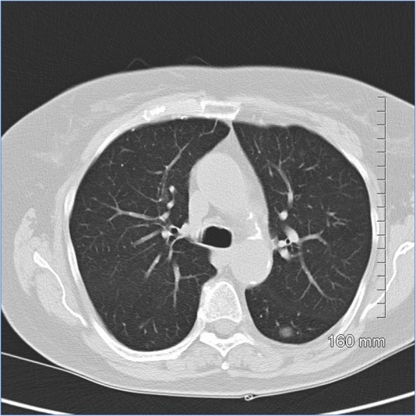
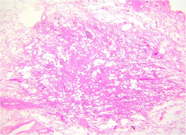
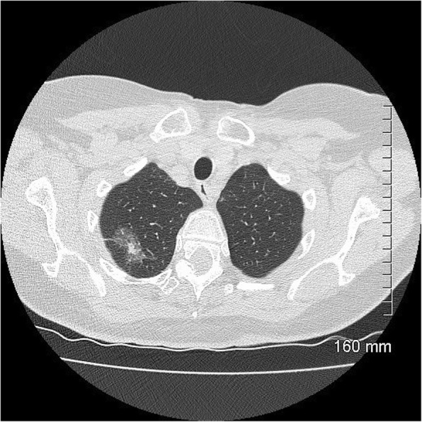
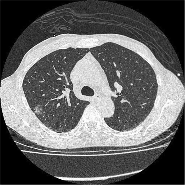
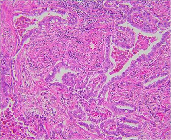
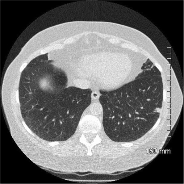
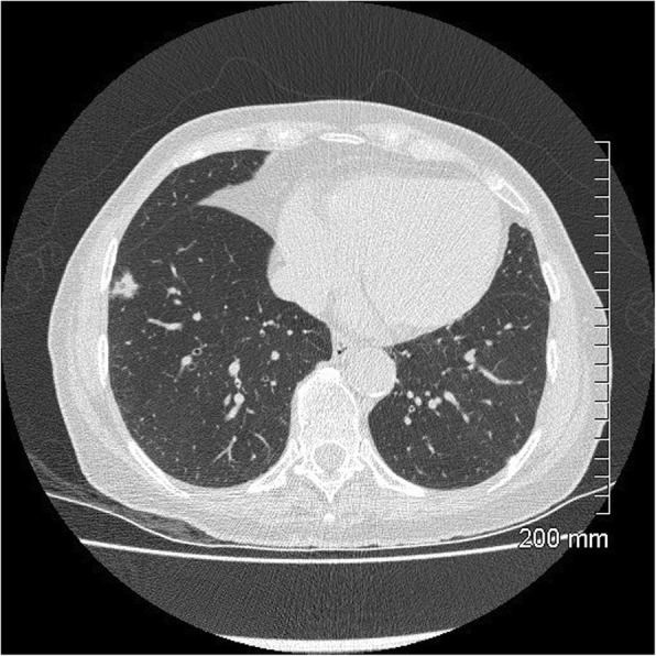
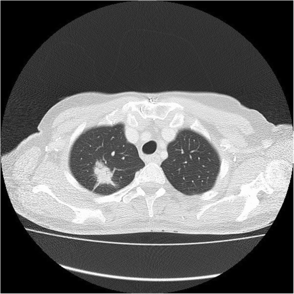
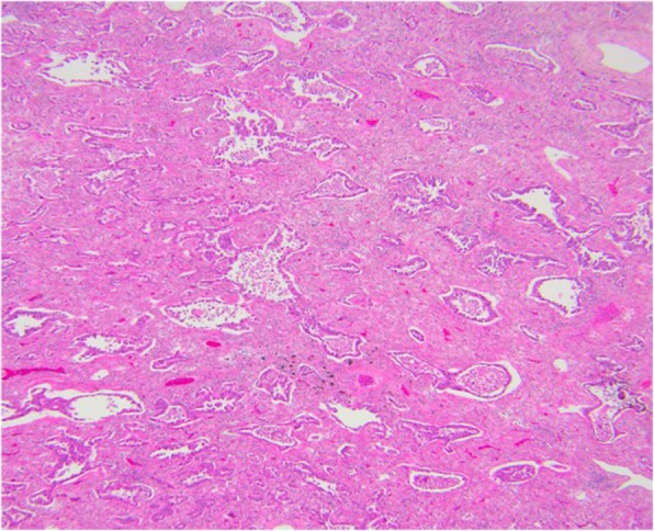
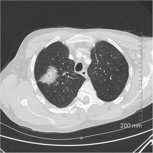
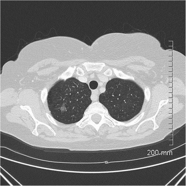
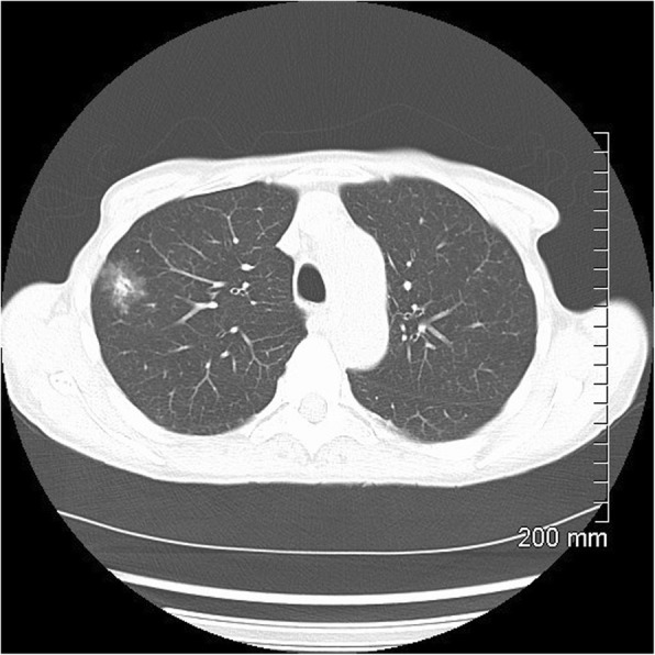
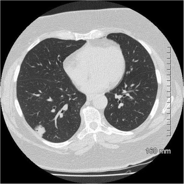
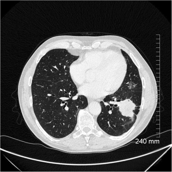
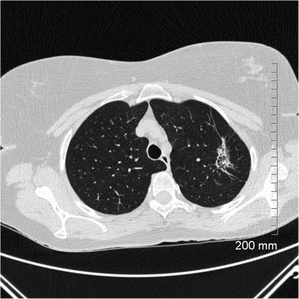
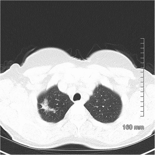
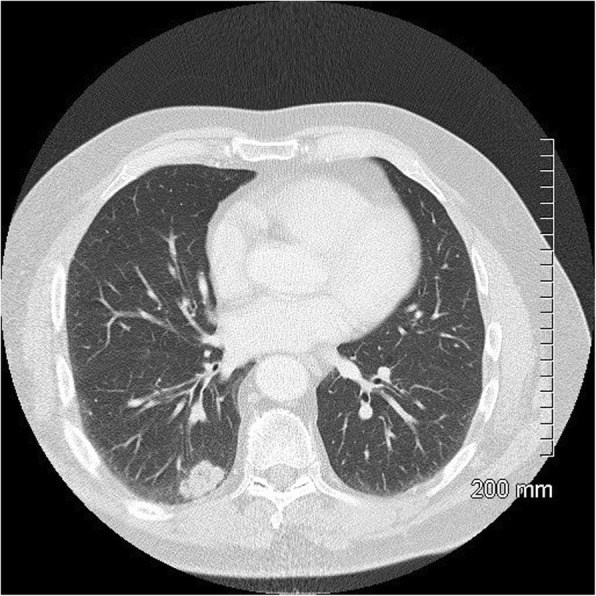
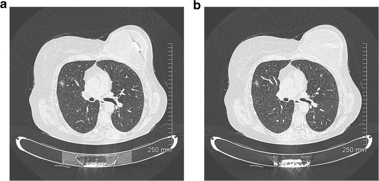
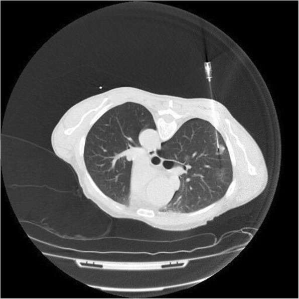
Adenocarcinoma in situ, minimally invasive adenocarcinoma, lepidic predominant adenocarcinoma and invasive mucinous adenocarcinoma are relatively new classification entities which replace the now retired term, bronchoalveolar carcinoma (BAC). The radiographic appearance of these lesions ranges from pure, ground glass nodules to large, solid masses. A thorough understanding of the new classification is essential to radiologists who work with MDT colleagues to provide accurate staging and treatment. A 2-year review was performed of all surgically resected cases of adenocarcinoma in situ, minimally invasive adenocarcinoma and lepidic predominant adenocarcinoma in our institution. Cases are broken down by age, gender, tumour type and tumour location. A pictorial review is presented to illustrate the radiologic and pathologic features of each entity.

Keywords: Computed tomography; Lung adenocarcinoma; Pathology; Solitary pulmonary nodule; Subsolid nodules.


Conflict of interest statement

Not applicable

Figures

Fig. 1
Fig. 1
AIS on CT. A 10-mm pure ground glass nodule in the left lower lobe. Stable in size for 3 years prior to biopsy
Fig. 2
Fig. 2
AIS histopathology: a 10-mm diameter adenocarcinoma pure lepidic growth, no invasion seen (H&E, 2 ×)
Fig. 3
Fig. 3
AIS on CT. A 33-mm ground glass opacity in the superior segment of the right upper lobe with a small, central solid component measuring 9 mm × 5 mm
Fig. 4
Fig. 4
MIA on CT. A 20-mm part-solid nodule, predominantly ground glass, in the right upper lobe
Fig. 5
Fig. 5
MIA histopathology: focal invasive growth < 5 mm diameter in a 17-mm lepidic pattern adenocarcinoma (H&E, 20 ×).
Fig. 6
Fig. 6
MIA on CT: 9 mm part-solid subpleural nodule, predominantly solid, in the left lower lobe
Fig. 7
Fig. 7
MIA on CT: 10 mm part-solid nodule, predominantly solid, in the right lower lobe
Fig. 8
Fig. 8
Lepidic predominant adenocarcinoma on CT: 3.2 cm predominantly solid mass in the right upper lobe with air bronchograms, thick spiculations and pleural tethering
Fig. 9
Fig. 9
Lepidic predominant adenocarcinoma histopathology: a 25-mm maximum diameter lepidic predominant adenocarcinoma where invasive acinar growth > 5 mm (H&E, 10 ×)
Fig. 10
Fig. 10
Lepidic predominant adenocarcinoma on CT. The 4.2 cm mass in the right upper lobe has spiculated margins, cystic lucencies, air bronchograms and satellite nodules
Fig. 11
Fig. 11
Favourable prognostic indicators: small size (< 2 cm). The pure ground glass nodule in the right upper lobe measures 10 mm. AIS
Fig. 12
Fig. 12
Favourable prognostic indicators: ground glass component. The part-solid nodule in the right upper lobe has a large ground glass component which correlates with lepidic growth. AIS
Fig. 13
Fig. 13
Favourable prognostic indicators: bubble-like lucencies in a part-solid right lower lobe nodule. MIA
Fig. 14
Fig. 14
Unfavourable prognostic indicators: thickened bronchovascular bundle. The 4-cm solid mass in the left lower lobe also demonstrates spiculated margins and pleural tethering with small foci of internal cavitation. A smaller ground glass nodule in the lingula was found to represent a synchronous AIS after surgical resection
Fig. 15
Fig. 15
Unfavourable prognostic indicators: coarse, thick spiculations. The area of mass-like consolidation in the left upper lobe also demonstrates cystic lucencies and air bronchograms. Lepidic predominant adenocarcinoma
Fig. 16
Fig. 16
Unfavourable prognostic indicators: large size (> 2 cm). The part-solid mass at the right apex measures 3.5 cm. MIA
Fig. 17
Fig. 17
Unfavourable prognostic indicators: concave notch. The solid, lobulated subpleural mass in the right lower lobe has a concave cut or notch on its medial border. Lepidic predominant adenocarcinoma
Fig. 18
Fig. 18
a, b Unfavourable prognostic indicators: focal pleural/fissural retraction. Part-solid nodule in the right upper lobe. Lepidic predominant adenocarcinoma
Fig. 19
Fig. 19
CT-guided biopsy of a part-solid nodule in the right upper lobe. Every effort should be made to target the solid component of the lesion which correlates with the invasive component of the nodule. The biopsy has been complicated by a small pneumothorax

References

    1. Travis WD, Garg K, Franklin WA, et al. Evolving concepts in the pathology and computed tomography imaging of lung adenocarcinoma and bronchioloalveolar carcinoma. J Clin Oncol. 2005;23(14):3279–3287. - PubMed
    1. Travis WD, Travis LB, Devesa SS. Lung cancer. Cancer. 1995;75(1 Suppl):191–202. - PubMed
    1. Malassez L (1876) Examen histologique d’un cas de cancer encephaloide du poumon (epithelioma). Arch Physiol Norm Pathol 1876;3:353-72 [Google Scholar].
    1. Musser JH (1903) Primary cancer of the lung. U Penn Med Bull 1903:16:289-96 [Google Scholar].
    1. Sweany HC (1935) A so-called alveolar cell cancer of the lung. Arch Pathol 1935;19:203-7 [Google Scholar].

LinkOut - more resources