Review
. 2020 May 19;11(1):67.
doi: 10.1186/s13244-020-00871-w.

Spectrum of imaging findings in AIDS-related diffuse large B cell lymphoma

Affiliations
Review

Spectrum of imaging findings in AIDS-related diffuse large B cell lymphoma

Edward Chege Nganga et al. Insights Imaging. .

Abstract

Lymphoma in HIV-infected patients is AIDS defining. This is the second most common AIDS defining malignancy after Kaposi's sarcoma. Development of lymphoma in HIV patients is related to immunosuppression and high viral load. Co-infection with other lymphotrophic viruses especially EBV is also strongly associated with development of lymphoma in HIV patients. Despite advances in HAART therapy, incidence of diffuse large B cell lymphoma in HIV-infected patients remains significantly higher than in the general population.Early diagnosis is challenging due to presence of opportunistic infections and atypical presentation of the lymphoma in this subset of patients. Atypical imaging findings are not unusual, and the diagnosis of lymphoma on imaging is on many occasions unexpected as the patient would ideally be initially investigated for presumed opportunistic infection.Lymphoma treatment approaches in HIV patients are complicated by comorbidity with opportunistic infections and performance status of the patients. Treatment failure and early relapse are also common in AIDS-related lymphoma. This review article highlights the common and unusual multimodality imaging findings in HIV-associated lymphoma.

Keywords: AIDS-related diffuse large B cell lymphoma; Extranodal lymphoma; HIV and malignancy; Multimodality imaging of lymphoma.


Conflict of interest statement

No competing interests

Figures

Fig. 1
Fig. 1
Chest radiograph of a 19-year-old HIV-positive patient with low CD4 count. He presented with cough and dyspnea and was initially being evaluated for possible atypical pneumonia. The chest radiograph showed multiple focal opacities predominantly in the lower lung zones. On CT, there were lung parenchymal nodules/masses of variable sizes which were also intensely FDG avid on PET CT. There was extensive lymphoma involvement elsewhere including in the liver, spleen (splenic activity higher than liver background), and multiple nodal sites above and below the diaphragm. Biopsy of the liver lesion was consistent with DLBCL
Fig. 2
Fig. 2
Coronal reformats of a CECT abdomen and pelvis of a recently diagnosed HIV-positive patient who presented with abdominal pain, diarrhea, and weight loss show long-segment bowel thickening involving the distal ileum and caecum (long arrow). There was also mesenteric lymphadenopathy, with some necrotic nodes, (short slender arrow) and a moderate ascites (short fat arrow). There were also enlarged centrally necrotic right inguinal lymph nodes
Fig. 3
Fig. 3
Axial CT images axial contrast-enhanced CT image and liver ultrasound of a patient with HIV-associated primary hepatic lymphoma. The contrast-enhanced CT shows a homogeneous soft tissue attenuation mass which does not show significant enhancement, and with fear preservation of the surrounding liver architecture. The mass displaces or encases the hepatic veins but does not invade vasculature or lead to tumor thrombus. On ultrasound, it appears as a fairly homogenous mass hypoechoic to the liver parenchyma. There is no increase vascularity within the mass or in its periphery
Fig. 4
Fig. 4
Coronal and axial CECT images of the abdomen (a and b) show hepatomegaly and a heterogenous hypoattenuating mass infiltrating through most of the right lobe of the liver. The infiltrative liver mass had more focal hypoattenuating areas (fat arrow). Aortocaval lymph nodes are also noted (short arrow). Axial CT through the pelvis in bone window (c) shows a permeative process involving the left femoral head and neck and associated pathological fracture
Fig. 5
Fig. 5
Ultrasound image of the left kidney in a HIV-positive male demonstrates heterogeneously echogenic areas in the medulla extending to the collecting system. Unenhanced CT KUB showed diffuse thickening of the renal pelvis with hyperdense soft tissue material relative to the renal parenchyma. Heterogenous infiltration of the kidney is seen on the postcontrast CT images in arterial and urographic phases
Fig. 6
Fig. 6
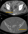
CECT of the pelvis in soft tissue window (a) and bone window (b). A mixed lytic and sclerotic process involving the sacrum and extending to the iliac bones through the sacroiliac joints is seen. There are associated pathological fractures and a large extra osseous soft tissue component. There are also enlarged necrotic bilateral pelvic sidewall and inguinal lymph nodes
Fig. 7
Fig. 7
A 46-year-old female patient who had pancreatic lymphoma (a) had a negative PET CT at the end of treatment (b and c). Nine months later, there was local disease recurrence at the pancreatic head on follow-up FDG PET CT scan (d and e)
Fig. 8
Fig. 8
Newly diagnosed HIV-positive patient who had CD4 count of 151 presented with primary gastric lymphoma. He was treated with R-CHOP regimen complicated by severe neutropenic sepsis. Intrathecal chemotherapy was deferred to cycle 3 due to poor performance. Interim PET/CT showed good treatment response of the gastric tumor but there was refractory neuraxial disease seen as FDG avid conus medullaris
Fig. 9
Fig. 9
This patient with extensive retroperitoneal and renal disease (a and b) showed excellent response to treatment in the interim PET CT (c, d, and e) after two cycles of R-CHOP. There was however early relapse with bulky disease developing in the retroperitoneum and the scrotum (f)
Fig. 10
Fig. 10
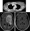
Axial CT image (a) demonstrates bilateral axillary lymphadenopathy, proven to be DLBCL after biopsy. The patient successfully completed treatment but developed CNS symptoms 1 year later. MRI of the brain (b) showed a large heterogenous mass in the left cerebral hemisphere which biopsy was proven to be lymphoma. A previous MRI brain (c) was negative for CNS involvement

References

    1. Global HIV & AIDS statistics — 2019 fact sheet. Available via: https://www.unaids.org/en/resources/fact-sheet.
    1. Kaplan LD, Ai W (2019) AIDS-related lymphomas: epidemiology, risk factors, and pathobiology. https://www.uptodate.com/contents/hiv-related-lymphomas-epidemiology-ris.... Accesed 14 Oct 2019.
    1. Javadi Sanaz, Menias Christine O., Karbasian Niloofar, Shaaban Akram, Shah Komal, Osman Adam, Jensen Corey T., Lubner Meghan G., Gaballah Ayman H., Elsayes Khaled M. HIV-related Malignancies and Mimics: Imaging Findings and Management. RadioGraphics. 2018;38(7):2051–2068. doi: 10.1148/rg.2018180149. - DOI - PubMed
    1. Carbone A, Gloghini A. AIDS-related lymphomas: from pathogenesis to pathology. Br J Haematol. 2005;130(5):662–670. doi: 10.1111/j.1365-2141.2005.05613.x. - DOI - PubMed
    1. Vishnu P, Aboulafia DM (2012) AIDS-related non-Hodgkin’s lymphoma in the era of highly active antiretroviral therapy. Adv Hematol 2012 - PMC - PubMed

LinkOut - more resources