Review
. 2020 Aug;145(2):83-96.
doi: 10.1007/s11120-020-00758-3. Epub 2020 May 19.

A comparative look at structural variation among RC-LH1 'Core' complexes present in anoxygenic phototrophic bacteria

Affiliations
Review

A comparative look at structural variation among RC-LH1 'Core' complexes present in anoxygenic phototrophic bacteria

Alastair T Gardiner et al. Photosynth Res. 2020 Aug.

Abstract

All purple photosynthetic bacteria contain RC-LH1 'Core' complexes. The structure of this complex from Rhodobacter sphaeroides, Rhodopseudomonas palustris and Thermochromatium tepidum has been solved using X-ray crystallography. Recently, the application of single particle cryo-EM has revolutionised structural biology and the structure of the RC-LH1 'Core' complex from Blastochloris viridis has been solved using this technique, as well as the complex from the non-purple Chloroflexi species, Roseiflexus castenholzii. It is apparent that these structures are variations on a theme, although with a greater degree of structural diversity within them than previously thought. Furthermore, it has recently been discovered that the only phototrophic representative from the phylum Gemmatimonadetes, Gemmatimonas phototrophica, also contains a RC-LH1 'Core' complex. At present only a low-resolution EM-projection map exists but this shows that the Gemmatimonas phototrophica complex contains a double LH1 ring. This short review compares these different structures and looks at the functional significance of these variations from two main standpoints: energy transfer and quinone exchange.

Keywords: Anoxygenic phototrophs; Light harvesting; Purple photosynthetic bacteria; RC–LH1; Reaction centres; Structures.


Conflict of interest statement

There are no conflicts of interest.

Figures

Fig. 1
Fig. 1
Two αβ-dimers in the ring of the light-harvesting complexes. Each view is slightly adjusted for maximal visibility. The Bchl tails and last few residues from some of the polypeptide chains have been omitted for clarity. The top row is in the plane of the membrane and bottom row is normal to the membrane. Common colours; α-polypeptides—purple blue, β-polypeptides—olive, ring Bchl—green, monomeric Bchl—lemon, carotenoids—orange, coordinating His residues—pink. a For comparison, a double αβ-dimer from the LH2 complex from Rbl. acidophilus. The monomeric Bchl is liganded to the C-terminal of the α-chain through a carboxy modified α-Met. The lower view is from the periplasmic side. b Blc. viridis with the additional γ-helix in yellow on the outside of the LH1 ring and between two β-helices. The lower view is from the cytoplasmic side. c Tch. tepidum with the co-ordinated Ca2+ on the cytoplasmic side shown in magenta and the water molecules as red spheres. This network of bonds requires residues from both αβ-dimers. The lower view is from the cytoplasmic side and part of the C-terminal α-polypeptide chain has been made partially transparent for clarity. d Rof. castenholzii with the monomeric Bchl liganded to a His residue on the α-polypeptide. The lower view is from the cytoplasmic side
Fig. 2
Fig. 2
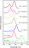
NIR absorption spectra of the RC–LH1 complexes mentioned in the text. The colour assignments are as follows along with the respective complex absorption band maxima (nm); a Rof. castenholzii, brown (800, 879), b Gem. phototrophica, pink (816, 868), c Rbl. acidophilus LH2, orange (804, 858), d Rba. sphaeroides, olive (873), e Tch. tepidum, blue (914), Rps. palustris, red (875), d Blc. viridis, green (1004)
Fig. 3
Fig. 3
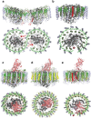
In plane (side on view) and membrane-normal (top side, periplasmic view) views of RC–LH1 complexes. The colour scheme is the same as in Fig. 1 with the fixed Cytochrome c given in salmon pink. a The dimeric complex from Rba. sphaeroides with gap-forming polypeptides PufX protein (red). b Rps. palustris with the gap-forming Protein W (red). c The LH1 ring from the complex in Tch. tepidum has no discernible gap and the ring of Ca2+ ions (magenta) is visible between each successive αβ-pair. d The Blc. viridis complex contains an additional smaller γ-polypeptide (yellow) that intercalates between the β-helices on the outside of the ring. The absence of this polypeptide at one site facilitates quinone/quinol exchange. e The Rof. castenholzii complex with the previously unknown, channel-forming subunit X (red). The channel is also formed by a transmembrane helix that protrudes down from the fixed Cytochrome c subunit. The enigmatic RC helix, TM7, mentioned in the text is represented in purple
Fig. 4
Fig. 4
Quinone/quinol exchange mechanisms used in the three high-resolution structures mentioned in the text. The colour scheme is the same as in Fig. 1, with the non-haem iron in firebrick red, QA quinone in blue, QB in cyan and any additional quinones resolved in the structures are in magenta. The top row presents a side view of the four αβ-helices in the complex where quinone/quinol exchange is presumed to take place. The bottom row is the equivalent membrane-normal view. Where required, a few of the terminal amino acids have been trimmed from the polypeptides termini in order to aid visibility. a Blc. viridis, the lower view is from the periplasmic side. b Tch. tepidum. The lower view shows the complete complex (minus the fixed cytochrome) from the cytoplasmic side with the same ring segment coloured that is presented above. The rest of the ring is given in grey and 80% transparency has been applied to the RC in order to help visualise QA and QB. c Rof. castenholzii has a relatively big gap produced by helix X (red) and the helix from the fixed Cytochrome (salmon pink). The lower view is from the periplasmic side

References

    1. Aagaard J, Sistrom WR. Control of synthesis of reaction center bacteriochlorophyll in photosynthetic bacteria. Photochem Photobiol. 1972;15:209. doi: 10.1111/j.1751-1097.1972.tb06240.x. - DOI - PubMed
    1. Barz WP, Francia F, Venturoli G, Melandri BA, Vermeglio A, Oesterhelt D. Role of PufX protein in photosynthetic growth of Rhodobacter sphaeroides 1 PufX is required for efficient light-driven electron transfer and photophosphorylation under anaerobic conditions. Biochemistry. 1995;34:15235–15247. doi: 10.1021/bi00046a032. - DOI - PubMed
    1. Barz WP, Oesterhelt D. Photosynthetic deficiency of a pufX deletion mutant of Rhodobacter sphaeroides is suppressed by point mutations in the light-harvesting complex genes pufB or pufA. Biochemistry. 1994;33:9741–9752. doi: 10.1021/bi00198a045. - DOI - PubMed
    1. Barz WP, Vermeglio A, Francia F, Venturoli G, Melandri BA, Oesterhelt D. Role of the PufX protein in photosynthetic growth of Rhodobacter sphaeroides. 2. PufX is required for efficient ubiquinone/ubiquinol exchange between the reaction center QB site and the cytochrome bc1 complex. Biochemistry. 1995;34:15248–15258. doi: 10.1021/bi00046a033. - DOI - PubMed
    1. Brunisholz RA, Jay F, Suter F, Zuber H. The light-harvesting polypeptides of Rhodopseudomonas viridis. The complete amino-acid sequences of B1015-alpha, B1015-beta and B1015-gamma. Biol Chem Hoppe-Seyler. 1985;366:87–98. doi: 10.1515/bchm3.1985.366.1.87. - DOI - PubMed

MeSH terms

Supplementary concepts

LinkOut - more resources