Review
. 2020 May 19;11(1):68.
doi: 10.1186/s13244-020-00874-7.

The role of contrast-enhanced ultrasound (CEUS) in the evaluation of scrotal trauma: a review

Affiliations
Review

The role of contrast-enhanced ultrasound (CEUS) in the evaluation of scrotal trauma: a review

Gibran T Yusuf et al. Insights Imaging. .

Abstract

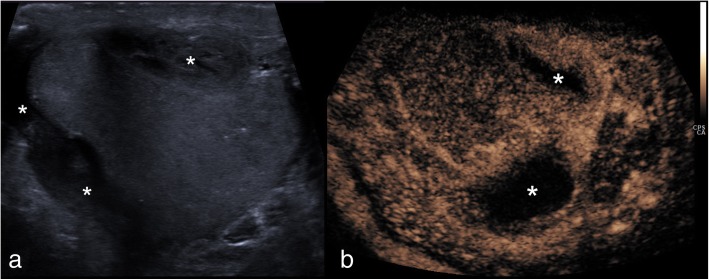
Testicular trauma is common, usually trivial and rarely requires hospital attendance, but if it does, then imaging becomes essential as scrotal assessment may be difficult due to pain and/or scrotal disruption. Ultrasound (US) assumes a crucial role as other cross-sectional modalities have a limited use in the acute presentation. Despite the acceptable accuracy of conventional US techniques, there are limitations which hinder a thorough evaluation, critically the assessment of tissue viability, crucial for clinical management and prognosis. Contrast-enhanced ultrasound (CEUS) has been shown to offer improved flow visualisation and tissue perfusion compared with conventional Doppler techniques. CEUS can accurately and confidently demonstrate the viability of testicular parenchyma, delineate fracture lines and haematomas and guide treatment for testis-sparing surgery or orchidectomy. The purpose of this review is to present the literature, familiarise physicians with the principles of CEUS and findings of scrotal trauma and illustrate the main abnormalities through characteristic and educative cases.

Keywords: Contrast-enhanced ultrasound; Haematoma; Scrotal trauma; Testicular trauma; Testis-sparing surgery.


Conflict of interest statement

PS has received lecture fees from Bracco, Siemens, Samsung, Philips and Hitachi. GTY has received lecture fees from Siemens and Bracco. MES has received lecture fees from Bracco. DYH has received lecture fees from Bracco. The rest of the authors have no conflict of interest regarding the publication of this manuscript.

Figures

Fig. 1
Fig. 1
Normal CEUS appearances of testicular parenchyma. Static image (a) showing the normal linear/striated mediastinal vessels outlined by microbubbles (arrowheads). Temporal prolonged contrast acquisition image (MIP) (b) showing the vascular architecture of normal testicular parenchyma
Fig. 2
Fig. 2
CEUS appearances of intratesticular haematoma after blunt trauma. B-mode image (a) showing an intratesticular rounded hypoechoic lesion (arrowheads), associated with hydrocele. On colour Doppler US (b), the lesion (arrowheads) showed no internal blood flow signals. CEUS (c) confirmed the absence of internal vascularity (asterisk), then unlikely a tumour and in keeping with that of haematoma. Note the peri-lesional hyperaemia (arrowheads) and the presence of two internal echoes, representing artefact from echogenic content
Fig. 3
Fig. 3
CEUS appearances of haematoma and ischaemia after blunt trauma. Dual-display image showing contrast-specific (left) and low MI B-mode image (right). A rounded hypoechoic and well-circumscribed lesion is seen (arrow), which shows no enhancement, in keeping with an intratesticular haematoma. Importantly, the lower pole of the testis appears heterogeneous on B-mode and shows no enhancement on CEUS, indicating the diagnosis of ischaemia (asterisk). The changes of the area of ischaemia are subtle on the B-mode image alone. Note the ability of CEUS to clearly define the borders of infarction which may be obscure on B-mode imaging alone
Fig. 4
Fig. 4
CEUS excluding ischaemia. B-mode (a) image demonstrated a hypoechoic, atrophic and heterogeneous lower pole (arrows) raising suspicion of post-surgical infarction. CEUS (b) demonstrated homogeneous perfusion of the entire testis, thus excluding the diagnosis and precluding the need for further surgical intervention
Fig. 5
Fig. 5
Conventional US and CEUS findings of testicular rupture. On B-mode (a), a hypoechoic linear disruption of the testicular parenchyma is demonstrated (arrowheads). Note the discontinuation of tunica albuginea with protrusion of echogenic material, indicating rupture (arrow). CEUS (b) determined the amount of viable parenchyma (arrowheads) which aided in pre-operative decision-making and allowed for the salvaging of viable testicular tissue avoiding the need for orchidectomy
Fig. 6
Fig. 6
CEUS findings of iatrogenic penetrating testicular injury post-biopsy. B-mode US image (a) obtained 24 h following core biopsy of the testis demonstrating two hypoechoic lesions within the central testis (arrowheads), which have typical appearances of intratesticular haematomas. CEUS (b) confirmed the avascular nature of the suspected haematomas (arrowheads). A separate patient with a histologically proven testicular tumour. For comparison, (c) B-mode shows a well-defined hypoechoic lesion (thick arrow). d CEUS demonstrated intrinsic enhancement (thin arrow) in keeping with a testicular tumour, unlike an avascular haematoma
Fig. 7
Fig. 7
CEUS findings of traumatic haematocele. B-mode US (a) showed an echogenic haematocele (asterisks) preventing assessment of the tunica albuginea contour on greyscale imaging. However, CEUS (b) clearly demonstrated normal testicular enhancement on both sides, without interruption of the tunica albuginea—excluding testicular infarction or rupture and establishing the diagnosis of a simple haematocele. Note that the haematocele (asterisks) shows no enhancement
Fig. 8
Fig. 8
CEUS appearances of extra-testicular haematoma. On B-mode (a), a note is made of an extratesticular hypoechoic haematoma (asterisks) causing compression of the testicular parenchyma. Essential issues in such cases include the thorough assessment of testicular parenchymal perfusion and integrity of the adjacent tunica, to rule our testicular rupture. CEUS (b) showed uniform enhancement of the testis and excludes focal ischaemia secondary to extrinsic compression, confirming that the tunica albuginea is intact. The extratesticular haematomas shows no internal enhancement (asterisks) but a rim of perilesional hyperaemia

References

    1. Lobianco R, Regine R, De Siero M, Catalano O, Caiazzo C, Ragozzino A. Contrast-enhanced sonography in blunt scrotal trauma. J Ultrasound. 2011;14:188–195. doi: 10.1016/j.jus.2011.09.003. - DOI - PMC - PubMed
    1. Yusuf GT, Sidhu PS. A review of ultrasound imaging in scrotal emergencies. J Ultrasound. 2013;16:171–178. doi: 10.1007/s40477-013-0033-x. - DOI - PMC - PubMed
    1. Lee SH, Lee DG, Choi SK, Choi T, Yoo KH. Trends in testicular injury in Korea, 1986-2015. J Korean Med Sci. 2017;32:1669–1673. doi: 10.3346/jkms.2017.32.10.1669. - DOI - PMC - PubMed
    1. Adlan T, Freeman SJ. Can ultrasound help to manage patients with scrotal trauma? Ultrasound. 2014;22:205–212. doi: 10.1177/1742271X14545911. - DOI - PMC - PubMed
    1. Sidhu Paul S. Clinical Ultrasound. 2011. Diseases of the testis and epididymis; pp. 593–620.

LinkOut - more resources