. 2020 Apr 30:14:1651-1662.
doi: 10.2147/DDDT.S249162. eCollection 2020.

Astaxanthin Attenuates Neuroinflammation in Status Epilepticus Rats by Regulating the ATP-P2X7R Signal

Affiliations

Astaxanthin Attenuates Neuroinflammation in Status Epilepticus Rats by Regulating the ATP-P2X7R Signal

Ming Wang et al. Drug Des Devel Ther. .

Abstract

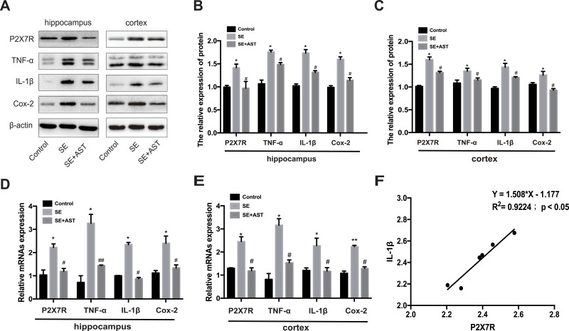
Background: As a life-threatening neurological emergency, status epilepticus (SE) is often refractory to available treatment. Current studies have shown a causal role of neuroinflammation in patients with lower seizure thresholds and driving seizures. The ATP-gated purinergic P2X7 receptor (P2X7R) is mainly expressed on the microglia, which function as gatekeepers of inflammation. Although emerging evidence has demonstrated significant anti-inflammatory effects of astaxanthin (AST) in SE, the associated mechanism remains unclear. Therefore, this study aimed to clarify the effects of AST on P2X7R-related inflammation in SE.

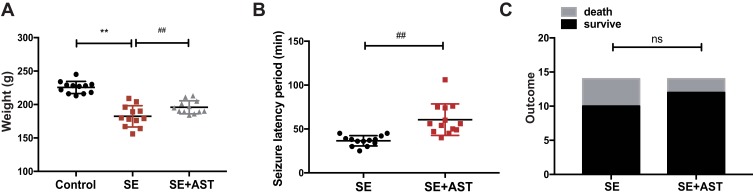
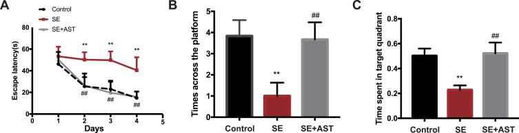
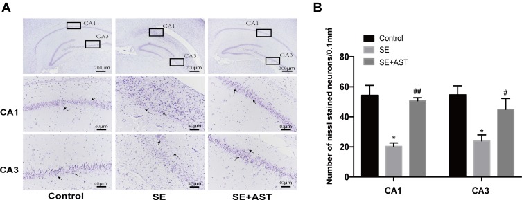
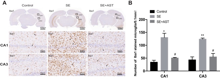
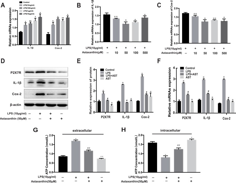
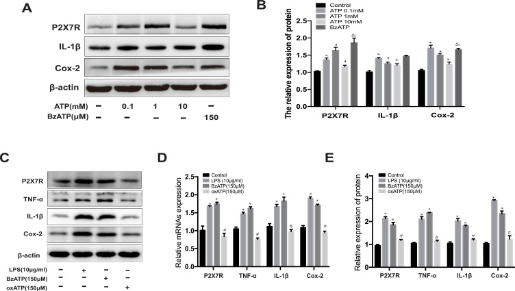
Methods: SE was induced in rats using lithium-pilocarpine, and AST was administered 1 h after SE induction. Rat microglia were treated with lipopolysaccharide (LPS), AST, ATP, 2,3-O-4-benzoyl-4-benzoyl-ATP (BzATP) and oxidized ATP (oxATP). The Morris water maze, immunohistochemistry, and Nissl staining were performed in rats. Expressions of P2X7R and inflammatory cytokines (such as cycloxygenase-2 (Cox-2), interleukin-1β (IL-1β), and tumor necrosis factor-α (TNF-α)) were detected using real-time polymerase chain reaction (RT-PCR) and Western blot (WB) both in rats and microglia. ATP concentration in the microglia was evaluated using ELISA.

Results: The AST alleviated hippocampal injury and improved cognitive dysfunction induced by SE. AST also effectively inhibited inflammation and downregulated P2X7R expression in both rat brain and microglia. The results also showed that AST reduced the extracellular ATP levels and that P2X7R expression could be increased by extracellular ATP. In addition, BzATP upregulates the expression of P2X7R and inflammatory factors in microglia. Conversely, it downregulates the expression of P2X7R and inflammatory factors.

Conclusion: Our study suggests that AST attenuated ATP-P2X7R mediated inflammation in SE.

Keywords: ATP; P2X7R; astaxanthin; neuroinflammation; status epilepticus.


Conflict of interest statement

The authors declare no conflict of interest.

Figures

Figure 1
Figure 1
Data from the rat assessed in study. (A) The weight of rats in control (n=12), SE (n=14), and SE+AST (n=14). (B) The seizure latency period to SE development in the SE and SE+AST group. (C) Rat survival after SE in SE and SE+AST group (**p <0.01 vs Control; ##p <0.01 vs SE). Abbreviation: ns, not significant.
Figure 2
Figure 2
The Morris water maze test in rats. (A) The escape latency of rats. (B) The time across the platform. (C) The spent time of rats in target quadrant (n=8, **p <0.01 vs Control; ##p <0.01 vs SE).
Figure 3
Figure 3
The Nissl stain of hippocampus in rats. (A) The Nissl stain of rat hippocampus. (B) The number of Nissl stained neurons. Left: control group. Middle: status epilepticus group. Right: astaxanthin (AST) treated group. The general morphology of the hippocampus in rat magnification 40×. The CA1 and CA3 regions of hippocampus are marked in squares. CA1 and CA3 region magnification 400×. Positively stained cells are marked with black arrows. The cell density per square millimeter was measured by ImageJ software (n=4, *p <0.05 vs Control; #p <0.05 vs SE; ##p <0.01 vs SE by one-way ANOVA).
Figure 4
Figure 4
The immunohistochemistry stain in rats. (A) The immunohistochemistry stained of Iba1 in rat hippocampus. (B) The number of stained microglia. Left: control group. Middle: status epilepticus group. Right: astaxanthin (AST) treated group. The general morphology of the hippocampus in rat magnification 20×. The CA1 and CA3 regions of hippocampus are marked in squares. CA1 and CA3 region magnification 400×. Positively stained cells are marked with black arrows (n=4, *p <0.05 vs Control; **p <0.01 vs Control; #p <0.05 vs SE by one-way ANOVA).
Figure 5
Figure 5
Effect of AST on P2X7R, IL-1β, Cox-2 and TNF-α expression in rats. (A) The protein P2X7R, TNF-a, IL-1β and Cox-2 levels in cortex and hippocampus. (B) The relative protein levels of P2X7R, TNF-α, IL-1β and Cox-2 in hippocampus. (C) The relative protein levels of P2X7R, TNF-α, IL-1β and Cox-2 in cortex. (D) The relative mRNAs levels of P2X7R, TNF-α, Cox-2 and IL-1β in the hippocampus. (E) The relative mRNAs levels of P2X7R, TNF-α, IL-1β and Cox-2 in the cortex. (F) Pearson correlation analysis of mRNA expression of P2X7R and IL-1β RNA in hippocampus of SE group. The relative expression of protein and mRNA were normalized to β-actin expression. Values are presented as the mean ± SD (n=5, *p < 0.05, **p < 0.01vs. Control; #p < 0.05, ##p < 0.01vs. SE).
Figure 6
Figure 6
Effect of astaxanthin on P2X7R, IL-1β, Cox-2 and ATP expression in LPS-induced rat microglia. (A) The mRNA expression of IL-1β and Cox-2 at different concentrations of LPS. (B, C) The mRNA expression of IL-1β and Cox-2 treated with different concentrations of AST. (D) WB analysis for P2X7R, IL-1β and Cox-2 protein levels in the rat microglia. (E) The expression levels of P2X7R, IL-1β and Cox-2 proteins in microglia. (F) The mRNA expression of P2X7R, IL-1β and Cox-2 were examined by RT-PCR. (G) The concentration of extracellular ATP (H) The concentration of intracellular ATP. The relative expression of protein and mRNA were normalized to β-actin expression. The concentration of extracellular and intracellular ATP was measured by ELISA kit. Cells were treated with designed concentration of AST (50μM) and LPS (10μg/mL) for 48h. Values are presented as the mean ± SD (*p < 0.05, **p < 0.01 vs Control group; #p < 0.05, ##p < 0.01 vs LPS group).
Figure 7
Figure 7
Effect of ATP on the P2X7R expression in microglia. (A) The protein expression of P2X7R, IL-1β and Cox-2 at different concentrations of ATP. (B) The relative protein of P2X7R, IL-1β and Cox-2. (C) Western blot analysis for P2X7R, IL-1β and Cox-2 protein levels in rat microglia. The expression level of P2X7R, TNF-α, IL-1β and Cox-2 proteins were normalized to β-actin expression. (D) The relative protein of P2X7R, IL-1β, TNF-α and Cox-2. (E) The relative mRNA expression of P2X7R, TNF-α, IL-1β and Cox-2 were analyzed by RT-PCR in rat microglia. Cells were treated ATP (0.1mM, 1mM and 10mM), LPS (10μg/mL), BzATP (150μM), and oxATP (150μM) for 48h in each group. The relative expression of protein and mRNA were normalized to β-actin expression. Values are presented as the mean ± SD (*p < 0.05 compared with the control group; #p < 0.05 compared with the BzATP group; &p < 0.05 compared with the ATP 1mM group).

Similar articles

Cited by

References

    1. Nelson SE, Varelas PN. Status epilepticus, refractory status epilepticus, and super-refractory status epilepticus. Continuum (Minneap Minn). 2018;24(6):1683–1707. doi:10.1212/CON.0000000000000668 - DOI - PubMed
    1. Lv RJ, Wang Q, Cui T, Zhu F, Shao XQ. Status epilepticus-related etiology, incidence and mortality: a meta-analysis. Epilepsy Res. 2017;136:12–17. doi:10.1016/j.eplepsyres.2017.07.006 - DOI - PubMed
    1. Trinka E, Kalviainen R. 25 years of advances in the definition, classification and treatment of status epilepticus. Seizure. 2017;44:65–73. doi:10.1016/j.seizure.2016.11.001 - DOI - PubMed
    1. Vezzani A, Dingledine R, Rossetti AO. Immunity and inflammation in status epilepticus and its sequelae: possibilities for therapeutic application. Expert Rev Neurother. 2015;15(9):1081–1092. doi:10.1586/14737175.2015.1079130 - DOI - PMC - PubMed
    1. Vezzani A, Friedman A, Dingledine RJ. The role of inflammation in epileptogenesis. Neuropharmacology. 2013;69:16–24. doi:10.1016/j.neuropharm.2012.04.004 - DOI - PMC - PubMed

MeSH terms