. 2020 Apr 17;7(Pt 3):509-521.
doi: 10.1107/S2052252520003917. eCollection 2020 May 1.

A structural study of TatD from Staphylococcus aureus elucidates a putative DNA-binding mode of a Mg2+-dependent nuclease

Affiliations

A structural study of TatD from Staphylococcus aureus elucidates a putative DNA-binding mode of a Mg2+-dependent nuclease

Kyu-Yeon Lee et al. IUCrJ. .

Abstract

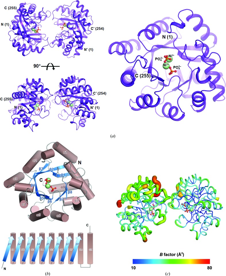
TatD has been thoroughly investigated as a DNA-repair enzyme and an apoptotic nuclease, and still-unknown TatD-related DNases are considered to play crucial cellular roles. However, studies of TatD from Gram-positive bacteria have been hindered by an absence of atomic detail and the resulting inability to determine function from structure. In this study, an X-ray crystal structure of SAV0491, which is the TatD enzyme from the Gram-positive bacterium Staphylococcus aureus (SaTatD), is reported at a high resolution of 1.85 Å with a detailed atomic description. Although SaTatD has the common TIM-barrel fold shared by most TatD-related homologs, and PDB entry 2gzx shares 100% sequence identity with SAV0491, the crystal structure of SaTatD revealed a unique binding mode of two phosphates interacting with two Ni2+ ions. Through a functional study, it was verified that SaTatD has Mg2+-dependent nuclease activity as a DNase and an RNase. In addition, structural comparison with TatD homologs and the identification of key residues contributing to the binding mode of Ni2+ ions and phosphates allowed mutational studies to be performed that revealed the catalytic mechanism of SaTatD. Among the key residues composing the active site, the acidic residues Glu92 and Glu202 had a critical impact on catalysis by SaTatD. Furthermore, based on the binding mode of the two phosphates and structural insights, a putative DNA-binding mode of SaTatD was proposed using in silico docking. Overall, these findings may serve as a good basis for understanding the relationship between the structure and function of TatD proteins from Gram-positive bacteria and may provide critical insights into the DNA-binding mode of SaTatD.

Keywords: DNA-binding protein; Staphylococcus aureus; TatD; X-ray crystallography; enzyme mechanisms; metal-dependent nuclease; protein structure; refinement; structure determination.


Figures

Figure 1
Figure 1
Overall structure of SaTatD. (a) The dimeric structure of SaTatD is presented as a ribbon diagram and colored purple. The Ni2+ ions are shown as spheres colored pale green; phosphates are shown in ball-and-stick representation with P and O atoms in orange and red, respectively. (b) The topology of the TIM-barrel fold constituting the SaTatD structure. Helices (brown) and strands (marine) are shown as cylindrical and ribbon diagrams, respectively. (c) B-factor distributions of SaTatD. The B-factor range (Å2) of the structure is shown as a gradient colored from deep blue to red. All of the structures were constructed using PyMOL (version 1.8; Schrödinger).
Figure 2
Figure 2
Distinct features of the SaTatD structure. (a) Chains A and B are shown as ribbon diagrams within the transparent surface colored purple and cyan, respectively. The dimeric interface areas consisting of Loop′1–Loop2–Loop3 (i) and Loop1–Loop′2–Loop′3 (ii) are expanded in the right panel. The residues contributing to hydrogen-bonding interactions are labeled and shown as lines. The interactions are presented as dotted lines (red). (b) LigPlot diagram presenting hydrophilic (red dotted lines) and hydrophobic (pink spoked curves) interactions in the dimeric interface. (c) The cis-peptide bonds observed in the SaTatD structure. Each cis-peptide is enlarged in the right panel (i, ii and iii) and shown as a red trapezoid. The related residues are shown in ball-and-stick representation with 2mF oDF c electron-density maps contoured at 2.0σ as a blue mesh.
Figure 3
Figure 3
Structural comparisons of SaTatD and its homologs. (a) Superimposed views of the monomers of SaTatD and its homologs identified by the DALI server: TatD-like DNase from S. aureus (strain MW2) (green; PDB entry 2gzx), TatD-like DNase from T. maritima (salmon; PDB entry 1j6o), the TatD–DNA complex from E. coli (red; PDB entry 4pe8), YcfH from E. coli (yellow; PDB entry 1yix) and YjjV from E. coli (lime; PDB entry 1zzm). All metals and ligands are shown as spheres and in ball-and-stick representation, respectively. The Ni2+ ion in PDB entry 2gzx and the Zn2+ ion in YcfH and YjjV are colored marine and orange, respectively. The C atoms of DNA (in PDB entry 4pe8) and the PEG ligand (in PDB entry 1zzm) are presented in ball-and-stick representation in black. (b) Multiple sequence alignment of homologs was performed using ClustalW and ESPript. The conserved residues and similar residues are presented in white on a red background and in red in blue boxes, respectively. The residues contributing to metal binding and catalysis are marked with green circles and blue stars, respectively.
Figure 4
Figure 4
The active site of SaTatD. (a) Electrostatic surface potential view of SaTatD. The acidic active-site pocket is marked with a white box, and basic (blue) and acidic (red) surfaces composed of Loop2–Loop3 and Loop1, respectively, are marked in dotted circles. (b) Metal-binding site of SaTatD. The key residues and two phosphates coordinating Ni2+ ions are labeled and shown as lines and in ball-and-stick representation, respectively. The interactions are shown as red dotted lines. The distance between the Ni2+ ions is labeled and is shown as a black dotted line. (c) The key residues contributing to the binding of phosphates are labeled and shown as lines. The distance between the P atoms of the phosphates is labeled and is shown as a black dotted line. (d) Superimposed view of SaTatD and the E. coli TatD–DNA complex (PDB entry 4pe8), revealing that the two phosphates of SaTatD resemble the scissile phosphates of DNA in E. coli TatD. T, thymine; C, cytosine; G, guanine. In (bd), 2mF oDF c electron-density maps contoured at 2.0σ are shown as a blue mesh.
Figure 5
Figure 5
Nuclease activity assay of SaTatD. (a) A metal-dependent DNase assay performed with 10 µM protein and 4 mM EDTA or 2 mM bivalent metals with 200 nM SaTatD genomic DNA (771 bp) as a substrate for 1 h at 37°C. Control groups (Con.) were prepared without SaTatD protein. (b) Protein or metal concentration-dependent DNase assay carried out with 2 mM Mg2+ or 4 µM protein, respectively, in the presence of 100 nM substrate for 1 h at 37°C. (c) Metal-dependent RNase assay. Fluorescence values (relative fluorescence units; RFU) were measured at Ex/Em = 490/520 nm for 1 h at 37°C. (d) DNase activity assay for the mutational study of SaTatD carried out with each mutant protein at 4 µM, 2 mM Mg2+, 200 nM SaTatD genomic DNA for 1 h at 37°C.
Figure 6
Figure 6
Proposed DNA-binding mode. (a) Prediction_1 for the DNA-binding mode of the SaTatD dimer with 18-mer dsDNA. Prediction_1 suggests that owing to the dimeric interface, DNA has difficulty approaching the active site of SaTatD. DNA is shown as a ribbon diagram, and the DNA strands are colored wheat (3′–5′) and dark gray (5′–3′). (b) Prediction_2 for the DNA-binding mode of the SaTatD monomer with 3-mer dsDNA considering structural hindrance. Elongated DNA chains that may bind to the basic surface area (blue dotted circle) consisting of Loop2–Loop3 are displayed as wheat and dark gray dotted lines.

References

    1. Biswas, L., Biswas, R., Nerz, C., Ohlsen, K., Schlag, M., Schäfer, T., Lamkemeyer, T., Ziebandt, A. K., Hantke, K., Rosenstein, R. & Götz, F. (2009). J. Bacteriol. 191, 5921–5929. - PMC - PubMed
    1. Brändén, C.-I. (1991). Curr. Opin. Struct. Biol. 1, 978–983.
    1. Brünger, A. T. (1992). Nature, 355, 472–475. - PubMed
    1. Chang, Z., Jiang, N., Zhang, Y., Lu, H., Yin, J., Wahlgren, M., Cheng, X., Cao, Y. & Chen, Q. (2016). Nat. Commun. 7, 11537. - PMC - PubMed
    1. Chen, L., Shen, D., Sun, N., Xu, J., Wang, W. & Dou, D. (2014). Mol. Plant Microbe Interact. 27, 1070–1080. - PubMed