Review
. 2020 May 5:8:314.
doi: 10.3389/fcell.2020.00314. eCollection 2020.

CD95 Structure, Aggregation and Cell Signaling

Affiliations
Review

CD95 Structure, Aggregation and Cell Signaling

Nicolas Levoin et al. Front Cell Dev Biol. .

Abstract

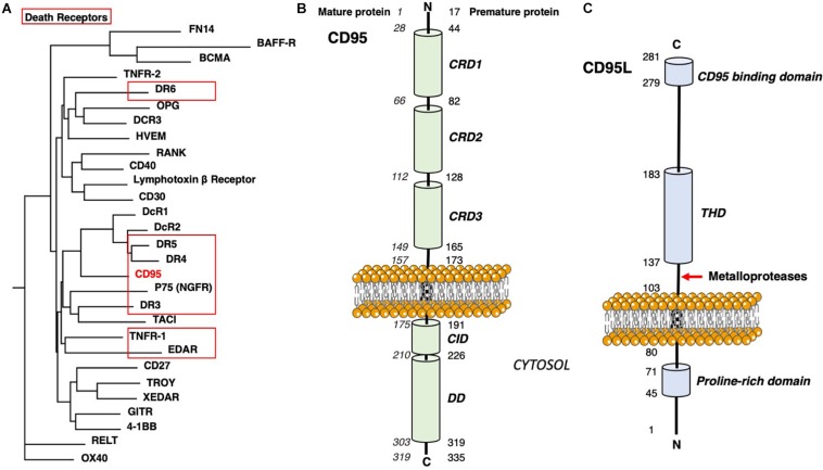
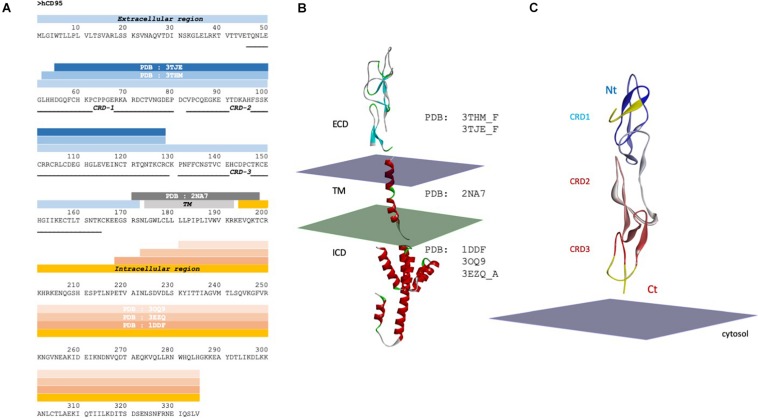
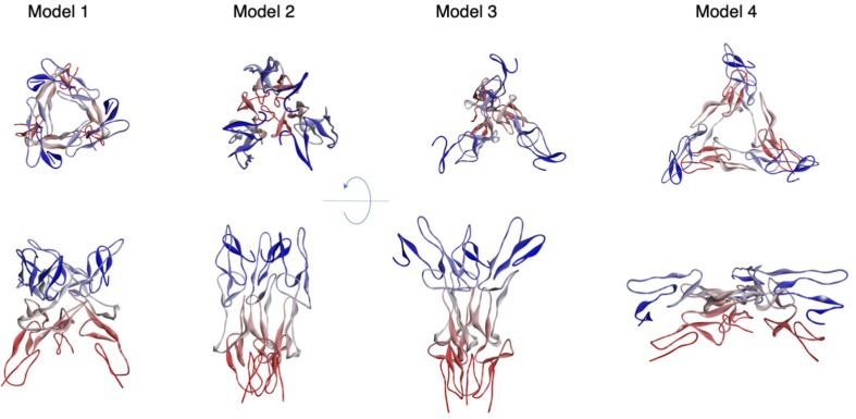
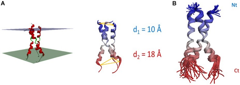
CD95 is a pre-ligand-associated transmembrane (TM) receptor. The interaction with its ligand CD95L brings to a next level its aggregation and triggers different signaling pathways, leading to cell motility, differentiation or cell death. This diversity of biological responses associated with a unique receptor devoid of enzymatic property raises the question of whether different ligands exist, or whether the fine-tuned control of CD95 aggregation and conformation, its distribution within certain plasma membrane sub-domains or the pattern of post-translational modifications account for this such broad-range of cell signaling. Herein, we review how the different domains of CD95 and their post-translational modifications or the different forms of CD95L can participate in the receptor aggregation and induction of cell signaling. Understanding how CD95 response goes from cell death to cell proliferation, differentiation and motility is a prerequisite to reveal novel therapeutic options to treat chronic inflammatory disorders and cancers.

Keywords: Fas; aggregation; apoptosis; inflammation; migration; stoichiometry.


Figures

FIGURE 1
FIGURE 1
CD95 domains. (A) Phylogenetic tree of human TNF receptors. DD-containing receptors are surrounded in red. Sequences were aligned with MAFFT 7 (Katoh and Standley, 2013) following L-INS-I strategy with BLOSUM 30 matrix, incorporating Mafft homologs and bootstrapping. MaxAlign option was used to increase the number of gap-free sites. The tree was built with Phylip 3.6 (Felsenstein, 1989) using a Neighbor-Joining method. (B) Main domains in the CD95 protein. (C) Main domains in the CD95L protein.
FIGURE 2
FIGURE 2
CD95 sequence and structure. (A) Sequence of CD95 with solved 3D structures and corresponding PDB ID code. Blue, gray and orange strips represent the extracellular domain, the transmembrane domain and the intracellular region of CD95, respectively. CRD, cysteine rich domain; TM, transmembrane; ICD, intracellular domain; ECD, extracellular domain. (B) Domains of a monomeric CD95 whose structure has been experimentally solved. The plasma membrane is symbolized by two parallel planes, with the outer leaflet in purple and the cytosolic couleur in green. Note that the orientation toward membrane is a hypothesis. (C) Structure of the extracellular domain of CD95. Crystal structure of CD95 ECD domain (PDB:3TJE), colored according to the sequence order (blue to red, from Nt to Ct extremities). The yellow structure (amino acid residues N31 to D55) represents the gap in the crystal structure, which has been completed using CD40 homology. Nt: Amino-terminal region; Ct: COOH-terminal region.
FIGURE 3
FIGURE 3
Models of trimeric CD95 ECD. Four models of the trimeric Apo CD95 ECD are depicted according to their decreasing docking score from left to right. Trimers were obtained by protomer docking, performed with SymmDock (Schneidman-Duhovny et al., 2005a). Ct ends are depicted in red, while the CD95 Nt region is in blue.
FIGURE 4
FIGURE 4
Structure and flexibility of CD95 TM. (A) Left panel: NMR-based structure of the trimeric TM helices according to PDB: 2NA7. The helix bundle is virtually inserted in a membrane, whose thickness is a hypothesis. Right panel: the Nt interdistance is less wide than its Ct counterpart (d1 = 9.7 ± 0.5 Å vs. d2 = 18.2 ± 2.6 Å between L174 or V195 Cα, for the 15 NMR structures). (B) Superposition of the 15 NMR structures, showing that the core of the bundle is quite rigid, while both ends are more flexible.
FIGURE 5
FIGURE 5
Structure and flexibility of CD95 ICD. (A) Superimposition of the two crystal structures of CD95 death domain (PDB:3EZQ in red and PDB:3OQ9 in yellow). In addition to the displacement of its juxtamembrane region, note the transformation of the α helices 5 and 6 within the death domain into a long stem helix. (B) Superimposition of Holo and Apo structures of the CD95 death domain (PDB:3OQ9 in yellow and PDB:1DDF in red). Note that there is still a conformational rearrangement of helices 5 and 6, but with a limited amplitude. (C) Different X-ray structures of ICD and their orientation toward the plasma membrane. Panels α to δ: proposed orientations of the tetrameric crystal structure of CD95:FADD complex (only CD95 is depicted). The N-terminal region of the death domain starting at N223 is the closest residue to TM and is labeled with black spheres. Chains in red seem correctly oriented regarding the plasma membrane, but the orientation of chains in yellow renders the position of the tetramer improbable. Panels γ to δ: only the closest dimers to the membrane are considered. Drawing in γ represents the most probable orientation toward the membrane. (D) Orientation of the pentameric CD95-DD, taking as reference the protomer showed in Figure 2B. Note that using this model, one DD is inserted into the plasma membrane. β. Optimized orientation of the pentameric CD95-DD regarding the position of the amino terminal residues K231 and Y232 (black balls and sticks) to the plasma membrane. Note the asymmetry of the structure, particularly for chain (A) (arrow).
FIGURE 6
FIGURE 6
CD95/CD95L complex and its association to the homotrimeric CD95 TM. (A) Manual alignment of trimeric Apo CD95 ECD model and experimental trimeric TM. Only residues E168GS are missing, and ECD E167 and TM R171 appeared close. (B) CD95-ECD was rebuilt using CD40 structure as a template. Next, this protomer was geometrically superimposed to the DcR3 homotrimer structure interacting with homotrimeric CD95L (yellow) with Maestro, Schrödinger Inc. (optimization of the α carbons superposition, with a final RMSD of 7.5 Å between DcR3 and CD95). CD95 TM is depicted in red. (C) Unlike the Apo CD95-ECD, the Holo Ct ends of CD95-ECD are too remote (each Ct end is distant from 40 Å) to be connected to the Nt ends of CD95-TM (in red) by the only 3 missing amino acids. The most probable rearrangement following ligand binding is a rotation/rocking of the whole CRD3 depicted in navy blue around N132 (the resulting modeled position is in navy blue ribbons, marked by blue arrows).

References

    1. Alderson M. R., Armitage R. J., Maraskovsky E., Tough T. W., Roux E., Schooley K., et al. (1993). Fas transduces activation signals in normal human T lymphocytes. J. Exp. Med. 178 2231–2235. 10.1084/jem.178.6.2231 - DOI - PMC - PubMed
    1. Anathy V., Aesif S. W., Guala A. S., Havermans M., Reynaert N. L., Ho Y. S., et al. (2009). Redox amplification of apoptosis by caspase-dependent cleavage of glutaredoxin 1 and S-glutathionylation of Fas. J. Cell Biol. 184 241–252. 10.1083/jcb.200807019 - DOI - PMC - PubMed
    1. Aoki K., Kurooka M., Chen J. J., Petryniak J., Nabel E. G., Nabel G. J. (2001). Extracellular matrix interacts with soluble CD95L: retention and enhancement of cytotoxicity. Nat. Immunol. 2 333–337. 10.1038/86336 - DOI - PubMed
    1. Arroyo-Manez P., Bikiel D. E., Boechi L., Capece L., Di Lella S., Estrin D. A., et al. (2011). Protein dynamics and ligand migration interplay as studied by computer simulation. Biochim. Biophys. Acta 1814 1054–1064. 10.1016/j.bbapap.2010.08.005 - DOI - PubMed
    1. Ashkenazi A., Dixit V. M. (1998). Death receptors: signaling and modulation. Science 281 1305–1308. 10.1126/science.281.5381.1305 - DOI - PubMed