. 2020 Jul 15;202(2):219-229.
doi: 10.1164/rccm.202003-0541OC.

Expression of the SARS-CoV-2 ACE2 Receptor in the Human Airway Epithelium

Affiliations

Expression of the SARS-CoV-2 ACE2 Receptor in the Human Airway Epithelium

Haijun Zhang et al. Am J Respir Crit Care Med. .

Abstract

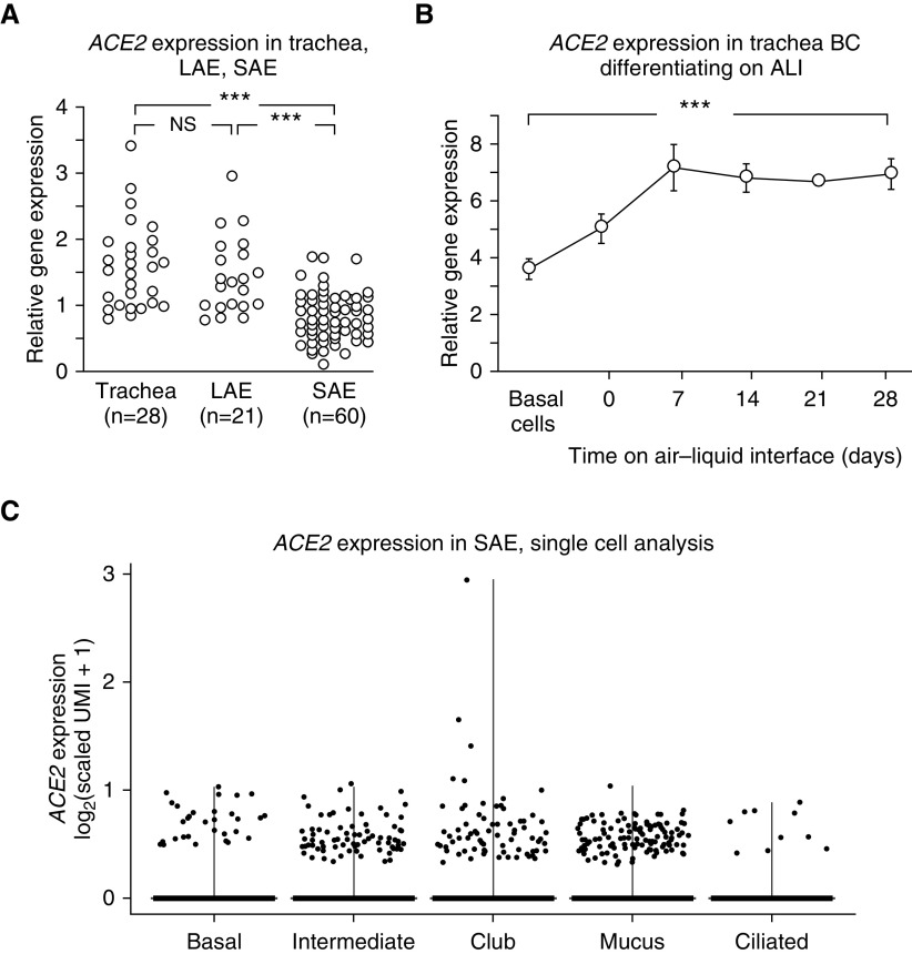
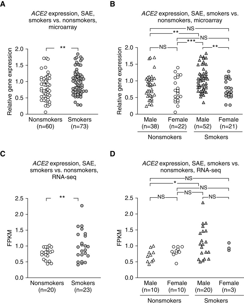
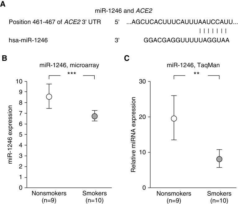
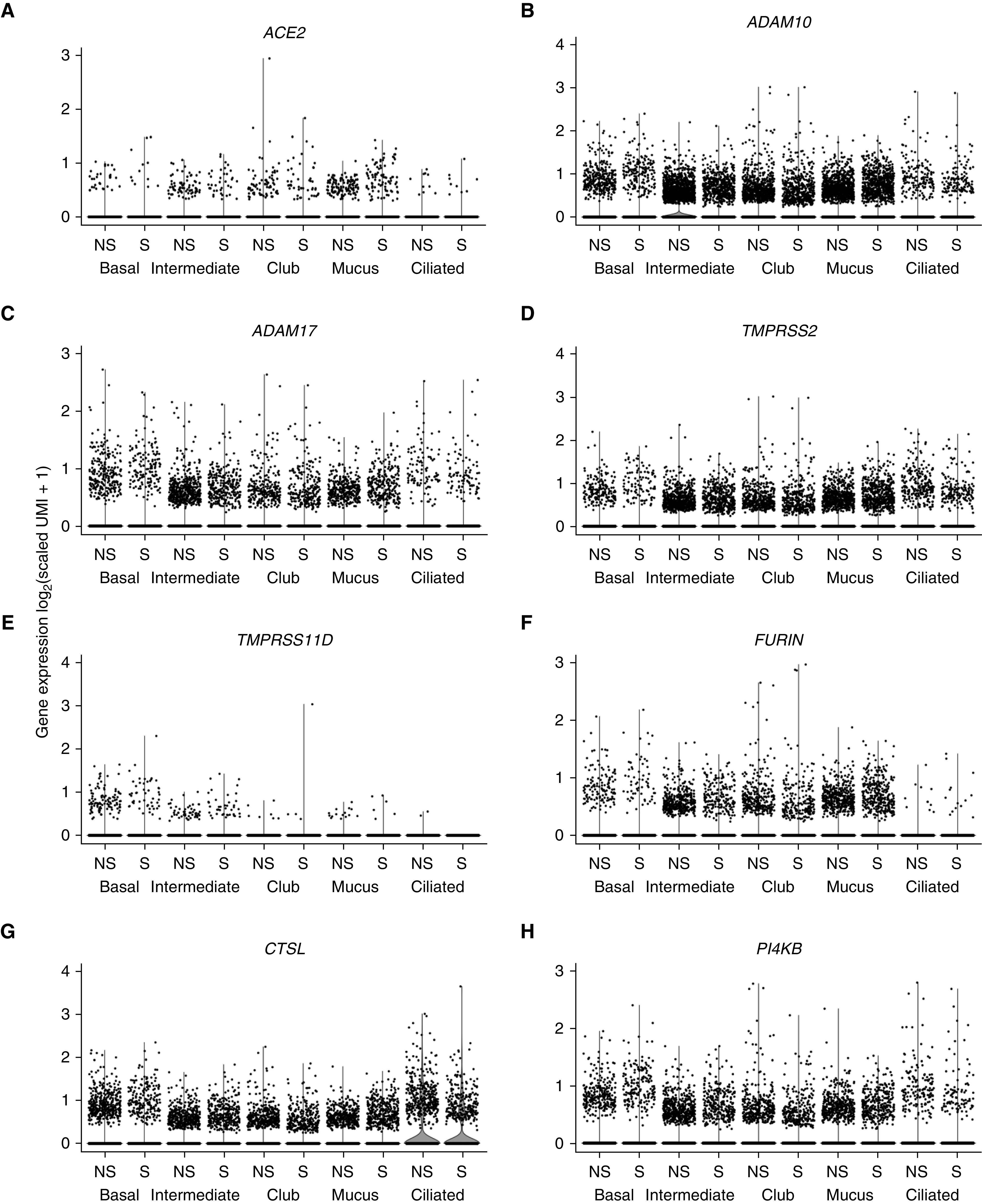
Rationale: Infection with the severe acute respiratory syndrome coronavirus 2 (SARS-CoV-2) causes coronavirus disease (COVID-19), a predominantly respiratory illness. The first step in SARS-CoV-2 infection is binding of the virus to ACE2 (angiotensin-converting enzyme 2) on the airway epithelium.Objectives: The objective was to gain insight into the expression of ACE2 in the human airway epithelium.Methods: Airway epithelia sampled by fiberoptic bronchoscopy of trachea, large airway epithelia (LAE), and small airway epithelia (SAE) of nonsmokers and smokers were analyzed for expression of ACE2 and other coronavirus infection-related genes using microarray, RNA sequencing, and 10x single-cell transcriptome analysis, with associated examination of ACE2-related microRNA.Measurements and Main Results:1) ACE2 is expressed similarly in the trachea and LAE, with lower expression in the SAE; 2) in the SAE, ACE2 is expressed in basal, intermediate, club, mucus, and ciliated cells; 3) ACE2 is upregulated in the SAE by smoking, significantly in men; 4) levels of miR-1246 expression could play a role in ACE2 upregulation in the SAE of smokers; and 5) ACE2 is expressed in airway epithelium differentiated in vitro on air-liquid interface cultures from primary airway basal stem/progenitor cells; this can be replicated using LAE and SAE immortalized basal cell lines derived from healthy nonsmokers.Conclusions:ACE2, the gene encoding the receptor for SARS-CoV-2, is expressed in the human airway epithelium, with variations in expression relevant to the biology of initial steps in SARS-CoV-2 infection.

Keywords: ACE2 transcriptome; COVID-19; coronavirus.


Figures

Figure 1.
Figure 1.
Expression of ACE2 (angiotensin-converting enzyme 2) in the human airway epithelium of healthy nonsmokers. (A and B) Expression level is presented as relative gene expression compared with all other genes on the array. See the online supplement for details on normalization. (A) Comparison of ACE2 expression in trachea epithelium, large airway epithelium, and small airway epithelium (SAE). Quantification was by Affymetrix HG-U133 Plus 2.0 microarrays. The data were generated from the data sets of Gene Expression Omnibus accession numbers 13933, 10135, and 11784 (23, 24, 26) and were compared using a two-way ANOVA (sex was identified as a source of variation). (B) ACE2 expression during in vitro differentiation of airway epithelium derived from primary tracheal basal cells (BCs). RNA was collected by brushing from freshly isolated, purified tracheal BCs and from cells derived from the BCs on an air–liquid interface culture at the initiation of the culture (Day 0) and at Days 7–28 of culture. ACE2 levels (determined by Affymetrix HG-U133 Plus 2.0 microarrays) increased as BCs differentiated into airway epithelial cells (ACE2 levels at Day 28 compared with Day 0, P < 10−5). (C) Single-cell 10x analysis of ACE2 expression in the different cell populations from the normal SAE of healthy nonsmokers. All the major cell types express ACE2, including basal, intermediate, club, mucus, and ciliated cells. Each data point represents a single cell. ACE2 was detected in a minority of epithelial cells from each cluster (detected in 1.2% of BCs, 2.6% of intermediate cells, 1.7% of club cells, 2.4% of mucus cells, and 1.0% of ciliated cells). These values are useful for comparison among the epithelial cell types but underestimate the actual percentage of cells expressing the gene (28). See the online supplement for markers used to define each cell type and for details on the calculation of scaled unique molecular identifiers and transformation for data presentation. ***P < 0.001. ALI = air–liquid interface; LAE = large airway epithelium; NS = nonsignificant; UMI = unique molecular identifiers.
Figure 2.
Figure 2.
Effect of smoking and sex on ACE2 (angiotensin-converting enzyme 2) expression in the small airway epithelium. (A and B) Expression level is presented as relative gene expression compared with all other genes on the array. See the online supplement for details on normalization. (A) Healthy smokers (gray symbols) versus nonsmokers (white symbols), with male and female sexes combined (Affymetrix HG-U133 Plus 2.0 microarrays). The data were generated from the data set of Tilley and colleagues (26), Gene Expression Omnibus accession number 11784. (B) Male sex versus female sex for smokers versus nonsmokers (Affymetrix HG-U133 Plus 2.0 microarrays), using the same data set as in A. (C) Smokers versus nonsmokers, male and female sexes combined, RNA sequencing (RNA-seq) (HiSeq 2500; Illumina). (D) Male versus female sex for smokers versus nonsmokers (RNA-seq), using the same data set as in C. A two-way ANOVA (sex was identified as a source of variation) was used for analysis. *P < 0.05, **P < 0.01, and ***P < 0.001. FPKM = fragments per kilobase of exon per million fragments sequenced; NS = nonsignificant; SAE = small airway epithelium.
Figure 3.
Figure 3.
Possible relationship of miR-1246 levels to modulate the levels of ACE2 (angiotensin-converting enzyme 2) in the small airway epithelium (SAE). Assessment of the data set of Wang and colleagues (27) (Gene Expression Omnibus accession number 53519) of healthy nonsmokers (n = 9; white circles) and healthy smokers (n = 10; gray circles) for smoking-related significant changes in levels of microRNA (miRNA) in the SAE for miR-1246 with sequences that complement the sequence of the 3′ untranslated region (3′UTR) of ACE2 mRNA. (A) Predicted pairing of target region in ACE2 3′UTR (top) and human miR-1246 (hsa-miR-1246, bottom) analyzed by TargetScanHuman 7.2 (http://www.targetscan.org/vert_72/). (B and C) miR-1246 levels are decreased in the SAE of smokers compared with those of nonsmokers. A two-way ANOVA (age was identified as a source of variation) was used for analysis. (B) Assessment by Affymetirx miRNA 2.0 arrays. Expression of miRNA is presented as relative miRNA expression compared with all other human mature miRNA. See online supplement for details. (C) Assessment by TaqMan PCR. Data are from Wang and colleagues (27). **P < 0.01 and ***P < 0.001.
Figure 4.
Figure 4.
Assessment of the small airway epithelia of healthy nonsmokers (n = 20; white circles) and smokers (n = 23; gray circles) for expression of genes that may be relevant to severe acute respiratory syndrome coronavirus 2 (SARS-CoV-2) infection. See text for details regarding the possible relevance of these genes to small airway epithelia infection by SARS-CoV-2. Quantification was by RNA sequencing (Illumina HiSeq 2500). A one-way ANOVA was used for analysis. (A) ADAM10. (B) ADAM17. (C) TMPRSS2. (D) TMPRSS11A. (E) TMPRSS11D. (F) FURIN. (G) CTSL. (H) PI4KB. **P < 0.01. FPKM = fragments per kilobase of exon per million fragments sequenced; NS = nonsignificant.
Figure 5.
Figure 5.
Single-cell 10x transcriptome analysis of the small airway epithelia of healthy nonsmokers (NS) (n = 5) and smokers (S) (n = 5) for expression of genes that may be relevant to severe acute respiratory syndrome coronavirus 2 (SARS-CoV-2) infection of the small airway epithelium. A total of 18,263 cells from NS and 16,678 cells from S were analyzed. ACE2 (angiotensin-converting enzyme 2) cells were detected in a minority of epithelial cells from both NS and S (percentage of cells in NS/percentage of cells in S: 1.2%/0.6% of basal cells, 2.6%/2.1% of intermediate cells, 1.7%/1.3% of club cells, 2.4%/1.9% of mucus cells, and 1.0%/1.2% of ciliated cells). These values are useful for comparison among the epithelial cell types but underestimate the actual percentage of cells expressing the gene (28). Expression of TMPRSS11A was detected by bulk RNA sequencing (HiSeq 2500; Illumina, Figure 4D) but was not detected by single-cell RNA sequencing (10x). For all comparisons of gene expression in all cell types, the differences in expression levels in NS and S were <10%; no significant differences in gene expression were observed. Statistical comparisons were performed using the Wilcoxon rank sum test, with P values adjusted using the Bonferroni correction. (A) ACE2. (B) ADAM10. (C) ADAM17. (D) TMPRSS2. (E) TMPRSS11D. (F) FURIN. (G) CTSL. (H) PI4KB. See the online supplement for markers used to define each cell type and for details on calculation of scaled unique molecular identifiers and transformation for data presentation. UMI = unique molecular identifiers.

Comment in

  • ACE2: The Only Thing That Matters?
    Sanchez-David RY, Swann OC, Peacock TP, Barclay WS. Sanchez-David RY, et al. Am J Respir Crit Care Med. 2020 Jul 15;202(2):161-163. doi: 10.1164/rccm.202006-2151ED. Am J Respir Crit Care Med. 2020. PMID: 32520592 Free PMC article. No abstract available.
  • Does Vaping Increase Susceptibility to COVID-19?
    Sharma P, Zeki AA. Sharma P, et al. Am J Respir Crit Care Med. 2020 Oct 1;202(7):1055-1056. doi: 10.1164/rccm.202005-2103LE. Am J Respir Crit Care Med. 2020. PMID: 32749868 Free PMC article. No abstract available.

References

    1. Chen N, Zhou M, Dong X, Qu J, Gong F, Han Y, et al. Epidemiological and clinical characteristics of 99 cases of 2019 novel coronavirus pneumonia in Wuhan, China: a descriptive study. Lancet. 2020;395:507–513. - PMC - PubMed
    1. Huang C, Wang Y, Li X, Ren L, Zhao J, Hu Y, et al. Clinical features of patients infected with 2019 novel coronavirus in Wuhan, China. Lancet. 2020;395:497–506. - PMC - PubMed
    1. Li Q, Guan X, Wu P, Wang X, Zhou L, Tong Y, et al. Early transmission dynamics in Wuhan, China, of novel coronavirus-infected pneumonia. N Engl J Med. 2020;382:1199–1207. - PMC - PubMed
    1. Wang D, Hu B, Hu C, Zhu F, Liu X, Zhang J, et al. Clinical characteristics of 138 hospitalized patients with 2019 novel coronavirus-infected pneumonia in Wuhan, China. JAMA. 2020;323:1061–1069. - PMC - PubMed
    1. Guan WJ, Ni ZY, Hu Y, Liang WH, Ou CQ, He JX, et al. China Medical Treatment Expert Group for Covid-19. Clinical characteristics of coronavirus disease 2019 in China. N Engl J Med. 2020;382:1708–1720. - PMC - PubMed

MeSH terms