. 2020 May 20;12(11):10457-10472.
doi: 10.18632/aging.103270. Epub 2020 May 20.

Ellagic acid protects mice against sleep deprivation-induced memory impairment and anxiety by inhibiting TLR4 and activating Nrf2

Affiliations

Ellagic acid protects mice against sleep deprivation-induced memory impairment and anxiety by inhibiting TLR4 and activating Nrf2

Wenjun Wang et al. Aging (Albany NY). .

Abstract

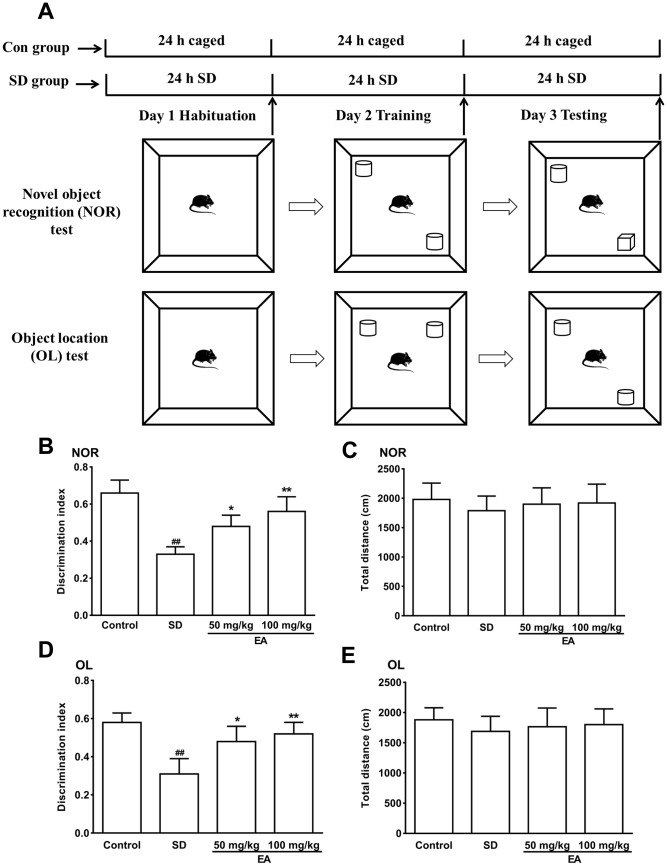
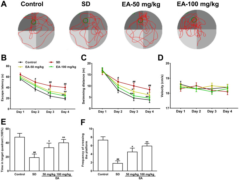
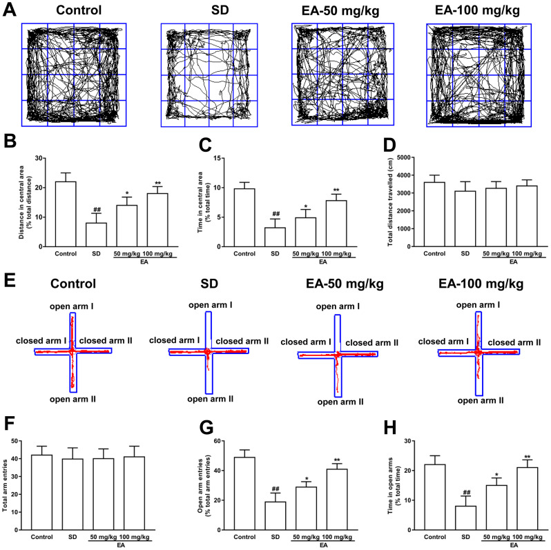
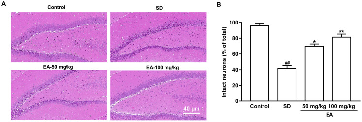
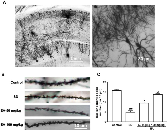
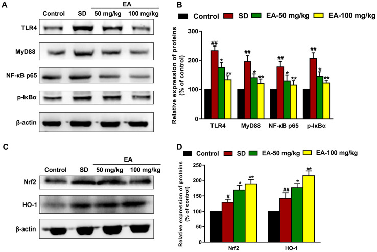
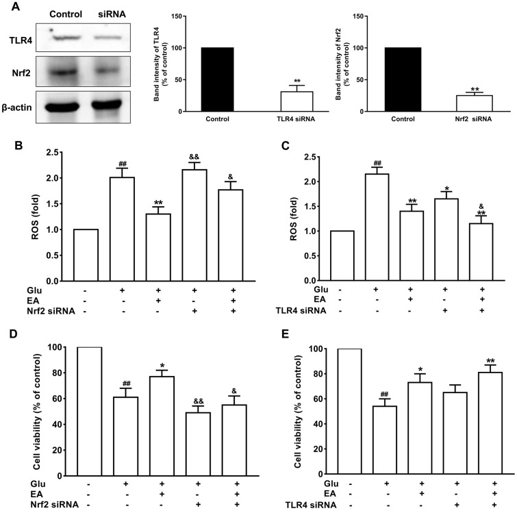
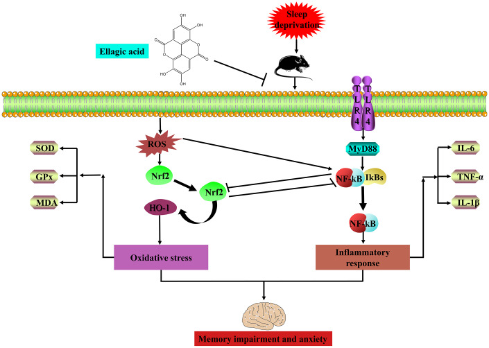
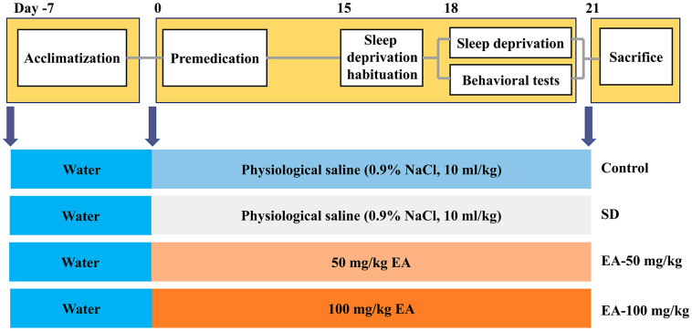
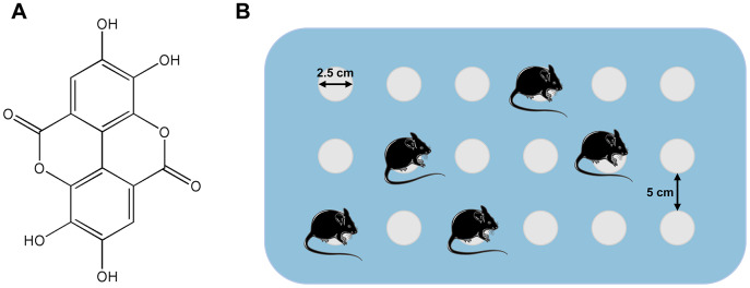
Sleep disorder has become a prevalent issue in current society and is connected with the deterioration of neurobehaviors such as mood, cognition and memory. Ellagic acid (EA) is a phenolic phytoconstituent extracted from grains and fruits that has potent neuroprotective properties. This research aimed to study the alleviative effect and mechanism of EA on memory impairment and anxiety caused by sleep deprivation (SD). EA ameliorated behavioral abnormalities in SD mice, associated with increased dendritic spine density, and reduced shrinkage and loss of hippocampal neurons. EA reduced the inflammatory response and oxidative stress injury caused by SD, which may be related to activation of the Nrf2/HO-1 pathway and mitigation of the TLR4-induced inflammatory response. In addition, EA significantly reduced the mortality and ROS levels in glutamate (Glu)-induced hippocampal neuron injury, and these effects of EA were enhanced in TLR4 siRNA-transfected neurons. However, knockdown of Nrf2 dramatically restrained the protective impact of EA on Glu-induced toxicity. Taken together, EA alleviated memory impairment and anxiety in sleep-deprived mice potentially by inhibiting TLR4 and activating Nrf2. Our findings suggested that EA may be a promising nutraceutical ingredient to prevent cognitive impairment and anxiety caused by sleep loss.

Keywords: Nrf2; TLR4; ellagic acid (EA); memory impairment; sleep deprivation (SD).


Conflict of interest statement

CONFLICTS OF INTEREST: The authors report no conflicts of interest in this work.

Figures

Figure 1
Figure 1
The novel object recognition (NOR) and object location test (OL) test performances were shown in AC. (A) Schematic of the NOR and OL tests. (B) Discrimination index toward a novel object and (C) total distance travelled (during 10 min test) were summarized. (D) The discrimination index toward a novel location and (E) total distance traveled (during the 10-minute test) were summarized. Data values were expressed as the mean ± SEM (n=12), ##P < 0.01 vs. control group; *P < 0.05 and **P < 0.01 vs. SD group.
Figure 2
Figure 2
Effect of CL on spatial reference memory in the MWM test in mice. (A) Representative swimming tracks in the MWM during the probe trial. (B) Mean daily escape latencies (time from the start to the hidden platform). (C) Distance travelled during the learning phase of the water maze task. (D) The swimming velocity of the mice. (E) The percentage of time spent in the target quadrant during the probe trial. (F) Frequency of crossing the target quadrant during the probe trials. All values were expressed as the mean ± SEM (n=12), #P < 0.05 and ##P < 0.01 vs. Control group; *P < 0.05 and **P < 0.01 vs. SD group.
Figure 3
Figure 3
Effect of CL on sleep deprivation induced anxiety-like behaviors. (A) Sample traces of locomotor activity in the open field test. (B) The total distance traveled and (C) time spent in the center area. (D) The total distance traveled (during the 15-minute test) was summarized. (E) Sample traces of locomotor activity in the elevated plus maze test. (F) The total arm entrances. (G) The entrance into the open arms and (H) time spent in the open arms. Data values were expressed as the mean ± SEM (n=12), ##P < 0.01 vs. control group; *P < 0.05 and **P < 0.01 vs. SD group.
Figure 4
Figure 4
EA improved neuronal survival after SD. (A) The hippocampus was stained by hematoxylin and eosin. (B) The percentage of intact neurons relative to the total neurons for each group (six different fields were counted per slice). Scale bar=40 μm. Data values were expressed as the mean ± SEM (n=3), #P < 0.05 and ##P < 0.01 vs. control group; *P < 0.05 and **P < 0.01 vs. SD group.
Figure 5
Figure 5
EA treatment reversed the spine density in the hippocampus area. (A) Golgi-Cox staining of CA1 pyramidal neurons for spine counting. (B) Representative images of basilar dendrites and (C) summary of spine counts from basilar dendrites. Data values were expressed as the mean ± SEM (n=3), ##P < 0.01 vs. control group; *P < 0.05 and **P < 0.01 vs. SD group.
Figure 6
Figure 6
EA modulated the Nrf2 and TLR4 signaling pathways. (A) and (C) The levels of Nrf2, HO-1, TLR4, MyD88, p-IκBα and NF-κB p65 in the hippocampus were detected by Western blot. (B) and (D) Band intensities were quantified as percentages of values from the control group. Data values were expressed as the mean ± SEM (n=3), #P < 0.05 and ##P < 0.01 vs. control group; *P < 0.05 and **P < 0.01 vs. SD group.
Figure 7
Figure 7
The protective effects of EA on glutamate-induced toxicity in neuronal cells. (A) The expression levels of Nrf2 and TLR4 significantly decreased in the siRNA treatment group. (B) and (C) Effect of EA on ROS levels in Nrf2 or TLR4 siRNA-transfected and Glu-treated neuronal cells. (D) and (E) Effect of EA on cell viability in Nrf2 or TLR4 siRNA-transfected and Glu-treated neuronal cells. Data values were expressed as the mean ± SEM (n=3), ##P < 0.01 vs. control group; *P < 0.05 and **P < 0.01 vs. Glu group; &P < 0.05 and &&P < 0.01 vs. EA-treated Glu group.
Figure 8
Figure 8
EA ameliorates sleep deprivation-induced memory impairment and anxiety via crosstalk between the Nrf2 and TLR4 pathways.
Figure 9
Figure 9
Experimental design procedure. Mice were randomly divided into four groups after habituation for 7 days. Then, mice were administered EA daily intraperitoneally EA for 21 days. After 3 days of SD habituation (from 8 a.m. to 11 a.m., 3 hours per day), all groups except the control group were subjected to SD for 72 hours (from 8 a.m. on day 18 to 8 a.m. on day 21). Behavioral tests were carried out after 24 hours of SD (Morris water maze training began on day 18).
Figure 10
Figure 10
(A) Structure of ellagic acids (PubChem CID: 5281855). (B) Simple illustration of the modified multiple-platform method used for SD mice.

References

    1. Gao T, Wang Z, Dong Y, Cao J, Lin R, Wang X, Yu Z, Chen Y. Role of melatonin in sleep deprivation-induced intestinal barrier dysfunction in mice. J Pineal Res. 2019; 67:e12574. 10.1111/jpi.12574 - DOI - PubMed
    1. Javaheripour N, Shahdipour N, Noori K, Zarei M, Camilleri JA, Laird AR, Fox PT, Eickhoff SB, Eickhoff CR, Rosenzweig I, Khazaie H, Tahmasian M. Functional brain alterations in acute sleep deprivation: an activation likelihood estimation meta-analysis. Sleep Med Rev. 2019; 46:64–73. 10.1016/j.smrv.2019.03.008 - DOI - PMC - PubMed
    1. Guo X, Keenan BT, Sarantopoulou D, Lim DC, Lian J, Grant GR, Pack AI. Age attenuates the transcriptional changes that occur with sleep in the medial prefrontal cortex. Aging Cell. 2019; 18:e13021. 10.1111/acel.13021 - DOI - PMC - PubMed
    1. Mullin BC, Phillips ML, Siegle GJ, Buysse DJ, Forbes EE, Franzen PL. Sleep deprivation amplifies striatal activation to monetary reward. Psychol Med. 2013; 43:2215–25. 10.1017/S0033291712002875 - DOI - PMC - PubMed
    1. McDonald DM, Keene AC. The sleep-feeding conflict: understanding behavioral integration through genetic analysis in drosophila. Aging (Albany NY). 2010; 2:519–22. 10.18632/aging.100181 - DOI - PMC - PubMed

Publication types

MeSH terms