. 2020 May 20;10(1):8377.
doi: 10.1038/s41598-020-65167-x.

The effect of wrist posture on extrinsic finger muscle activity during single joint movements

Affiliations

The effect of wrist posture on extrinsic finger muscle activity during single joint movements

Carl R Beringer 3rd et al. Sci Rep. .

Abstract

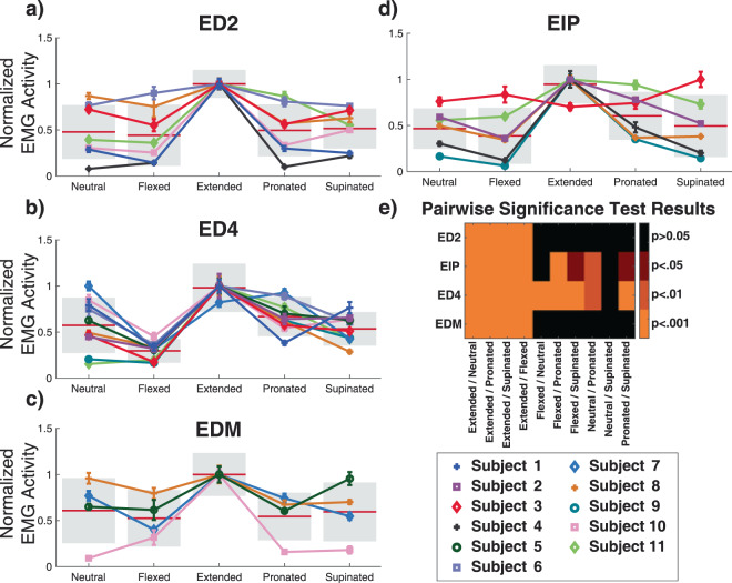
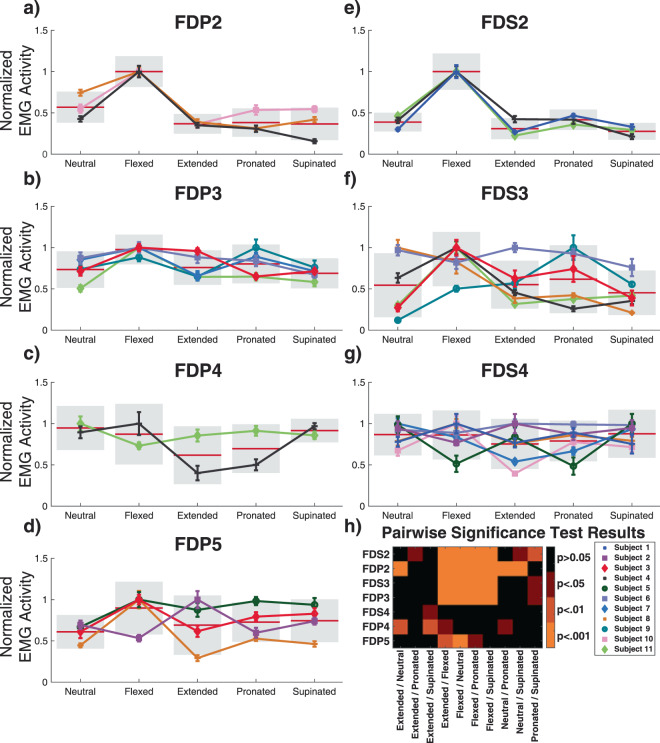
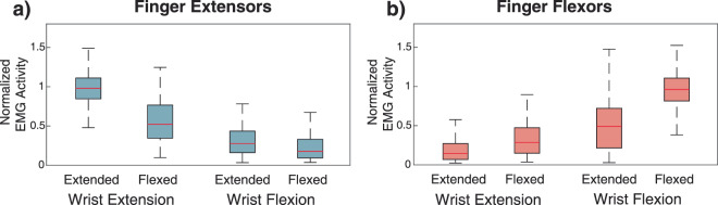
Wrist posture impacts the muscle lengths and moment arms of the extrinsic finger muscles that cross the wrist. As a result, the electromyographic (EMG) activity associated with digit movement at different wrist postures must also change. We sought to quantify the posture-dependence of extrinsic finger muscle activity using bipolar fine-wire electrodes inserted into the extrinsic finger muscles of able-bodied subjects during unrestricted wrist and finger movements across the entire range of motion. EMG activity of all the recorded finger muscles were significantly different (p < 0.05, ANOVA) when performing the same digit movement in five different wrist postures. Depending on the wrist posture, EMG activity changed by up to 70% in individual finger muscles for the same movement, with the highest levels of activity observed in finger extensors when the wrist was extended. Similarly, finger flexors were most active when the wrist was flexed. For the finger flexors, EMG variations with wrist posture were most prominent for index finger muscles, while the EMG activity of all finger extensor muscles were modulated in a similar way across all digits. In addition to comprehensively quantifying the effect of wrist posture on extrinsic finger EMG activity in able-bodied subjects, these results may contribute to designing control algorithms for myoelectric prosthetic hands in the future.


Conflict of interest statement

The authors declare no competing interests.

Figures

Figure 1
Figure 1
Overview of the experimental setup. (a) Electromagnetic tracking glove that subjects wore during the experiments. The red rectangles show placement of electromagnetic sensors in relation to the joints of the fingers. (b) Subjects viewed and followed a video which demonstrated the hand posture, movement, and timing.
Figure 2
Figure 2
Overview of data processing and effects of wrist posture on EMG activity. (a) Comparison of EMG activity during rest before (left) and after (right) electromagnetic noise removal. Blocked in red are examples of electromagnetic artifacts introduced by the kinematic tracking system and the same signal after electromagnetic artifact removal. Left: EMG activity after high-pass filtering to remove DC offset and motion artifacts. The electromagnetic artifacts can be observed boxed in red. Right: The same EMG activity after electromagnetic noise removal. (b) D2 metacarpal-phalangeal joint velocity and EMG activity of EIP and FDS2 during repeated flexion (red) and extension (blue) movements. The unshaded region between movements represents brief holding periods that were not included in calculations of EMG activity. (c) Rectified and processed EMG activity of ED4 during repetitions of D4 flexion and extension in neutral, flexed, extended, pronated, and supinated postures. The dashed vertical line shows the task start cue. The hand was held in these static postures for approximately 4 seconds before performing 10 movement repetitions. Note the substantial change in EMG activity for the same finger movement in different wrist postures.
Figure 3
Figure 3
(a–d) Overall mean normalized EMG activity for each subject for the extrinsic finger extensors during extension motions with the wrist in neutral, flexed, extended, pronated, and supinated postures. All finger extensors had their maximum EMG activity during extension movements when the wrist was held extended. Individual data points represent the subject mean and error bars are standard error of the mean. Data are color and marker coded for each subject. The horizontal red bars at each wrist posture show normalized group means and the standard deviation across subjects is shown as a gray box. Pairwise comparisons of EMG activity between the wrist extended and all other postures showed significant differences (p < 0.001, Tukey’s HSD). (e) Heat map of pairwise significant differences for EMG activity of the extrinsic finger extensor muscle for all combinations of wrist postures.
Figure 4
Figure 4
(a–g) Overall mean normalized EMG activity for each subject for the extrinsic finger flexors during flexion motions with the wrist in neutral, flexed, extended, pronated, and supinated postures. The D2, D3, and D5 finger flexors showed significantly elevated EMG activity when the wrist was flexed. Individual data points represent the subject mean and standard error of the mean. Data are color and marker coded for each subject. The horizontal red bars show normalized group means and the standard deviation is shown as a gray box. (h) Heat map of pairwise significant differences (Tukey’s HSD) of EMG activity between postures for the extrinsic finger flexors.
Figure 5
Figure 5
Normalized mean EMG activity for (a) finger extensors (N = 22 muscles) and (b) finger flexors (N = 28 muscles) during wrist extension and flexion movements in flat and fist postures. For each of the 11 subjects, 10 repetitions were performed for movements in each posture. The red line shows the median, and the outer boxes are the first and third quartile. Error bars represent the 5–95% confidence interval. All posture and movement combination showed significant differences (p < 0.005, Kruskal-Wallis with Dunn post-hoc testing).

References

    1. An KN, Hui FC, Morrey BF, Linscheid RL, Chao EY. Muscles across the elbow joint: A biomechanical analysis. J. Biomech. 2017;14:659–661. doi: 10.1016/0021-9290(81)90048-8. - DOI - PubMed
    1. Li Z-M, Latash ML, Zatsiorsky VM. Force sharing among fingers as a model of the redundancy problem. Exp. brain Res. 1998;119:276–286. doi: 10.1007/s002210050343. - DOI - PubMed
    1. Zatsiorsky, V. M., Li, Z.-M. & Latash, M. L. Enslaving effects in multi-finger force production. Exp. brain Res. 131, (2000). - PubMed
    1. Schieber MH, Gardinier J, Liu J. Tension distribution to the five digits of the hand by neuromuscular compartments in the macaque flexor digitorum profundus. J. Neurosci. 2001;21:2150–2158. doi: 10.1523/JNEUROSCI.21-06-02150.2001. - DOI - PMC - PubMed
    1. Leijnse JNAL, Campbell-Kyureghyan NH, Spektor D, Quesada PM. Assessment of individual finger muscle activity in the extensor digitorum communis by surface EMG. J. Neurophysiol. 2008;100:3225–3235. doi: 10.1152/jn.90570.2008. - DOI - PubMed

Publication types