. 2020 May 20;10(1):8323.
doi: 10.1038/s41598-020-65155-1.

PGAM5-MAVS interaction regulates TBK1/ IRF3 dependent antiviral responses

Affiliations

PGAM5-MAVS interaction regulates TBK1/ IRF3 dependent antiviral responses

Yu-Qiang Yu et al. Sci Rep. .

Abstract

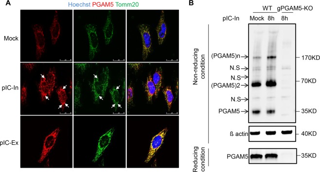
Viral infections trigger host innate immune responses, characterized by the production of type-I interferons (IFN) including IFNβ. IFNβ induces cellular antiviral defense mechanisms and thereby contributes to pathogen clearance. Accumulating evidence suggests that mitochondria constitute a crucial platform for the induction of antiviral immunity. Here we demonstrate that the mitochondrial protein phosphoglycerate mutase family member 5 (PGAM5) is important for the antiviral cellular response. Following challenge of HeLa cells with the dsRNA-analog poly(I:C), PGAM5 oligomers and high levels of PGAM5 were found in mitochondrial aggregates. Using immunoprecipitation, a direct interaction of PGAM5 with the mitochondrial antiviral-signaling protein (MAVS) was demonstrated. In addition, PGAM5 deficient cells showed diminished expression of IFNβ and IFNβ target genes as compared to WT cells. Moreover, PGAM5 deficient mouse embryonic fibroblasts (MEFs) exhibited decreased phosphorylation levels of IRF3 and TBK1 when challenged with poly(I:C) intracellularly. Finally, PGAM5 deficient MEFs, upon infection with vesicular stomatitis virus (VSV), revealed diminished IFNβ expression and increased VSV replication. Collectively, our study highlights PGAM5 as an important regulator for IFNβ production mediated via the TBK1/IRF3 signaling pathway in response to viral infection.


Conflict of interest statement

The authors declare no competing interests.

Figures

Figure 1
Figure 1
Intracellular poly(I:C) induces the formation of PGAM5 multimers. (A) Confocal microscopy imaging of HeLa cells stimulated with 50 µg/ml extracellular poly(I:C) (pIC-Ex) or 1 µg/ml intracellular poly(I:C) (pIC-In) for 8 h and stained with the following antibodies: anti-Tomm20 and anti-PGAM5. Hoechst was used to stain the nucleus. Extracellular poly(I:C) was added directly into the medium. Intracellular poly(I:C) was transfected into the cells. Arrowheads indicate PGAM5 aggregates. (B) SDS-PAGE analysis of PGAM5 multimers in lysates of HeLa cells under non-reducing or reducing conditions. HeLa cells were stimulated with 1 µg/ml intracellular poly(I:C) for 8 h. Cell lysates of PGAM5 CRISPR/Cas9 knockout cells were used as control to exclude non-specific bands. N.S, non-specific band.
Figure 2
Figure 2
PGAM5 regulates intracellular poly(I:C)-induced IFNβ expression. (A) Immunoblot analysis of PGAM5 in lysates of WT and PGAM5 CRISPR/Cas9 knockout HeLa cells. β-actin served as a loading control. (B) mRNA expression of IFNB in WT and PGAM5 CRISPR/Cas9 knockout HeLa cells treated with vehicle (Mock), 1 µg/ml intracellular poly(I:C) (p.IC-In), 50 µg/ml extracellular poly(I:C) (p.IC-Ex), 1 µg/ml intracellular poly(dI:dC), or 100 ng/ml LPS. N.S., not significant. (C) mRNA expression of IFI27 and CXCL10 in WT and PGAM5 CRISPR/Cas9 knockout HeLa cells stimulated with 1 µg/ml intracellular poly(I:C) for 8 h. (D) Immunoblot analysis of PGAM5 in lysates of WT and PGAM5−/− Mouse Embryonic Fibroblasts (MEFs). β-actin served as a loading control. (E,F) mRNA expression of Ifnb, Ifit1 and Il6 in WT and PGAM5−/− MEFs stimulated with 1 µg/ml intracellular poly(I:C) for 8 h. (G) Immunoblot of ectopic PGAM5 expression in lysates of PGAM5 CRISPR/Cas9 knockout HeLa cells transfected for 48 h with full-length PGAM5 (P) vectors or phosphatase mutant PGAM5 (P105A) vectors. Lysates from empty vector transfected WT and PGAM5 CRISPR/Cas9 knockout HeLa cells were used as control. (H) mRNA expression of IFNB in corresponding HeLa cells stimulated with or without intracellular poly(I:C) for 8 h. WT + Flag, WT cells transfected with empty vector. gPGAM5-KO + Flag, PGAM5 CRISPR/Cas9 knockout cells transfected with empty vector. gPGAM5-KO + Flag PGAM5, PGAM5 CRISPR/Cas9 knockout cells transfected with full-length PGAM5 vectors. gPGAM5-KO + Flag PGAM5-105A, PGAM5 CRISPR/Cas9 knockout cells transfected with phosphatase mutant PGAM5 vectors. Experiments were performed three times and representative data are shown. Data are presented as mean +SD and student’s t-test was used for statistical calculation. **P < 0.01 and ***P < 0.001.
Figure 3
Figure 3
PGAM5 interacts with MAVS by regulating TBK1/IRF3 dependent IFNβ expression. (A) Immunoblot of phosphorylated or total proteins in lysates of WT and PGAM5−/− MEFs stimulated with intracellular poly(I:C) at different time points. (B) Relative analysis of pTBK1 (left panel) and pIRF3 (right panel) intensity. The total protein level of IRF3 or TBK1 was used for normalization. (C) mRNA expression of Ifnb in MEFs (left panel) or IFNB in HeLa cells (right panel) stimulated with intracellular poly(I:C) in the presence or absence of TBK1 inhibitor BX795. DMSO was used as control treatment. Experiments were performed three times and representative data are shown. Data are presented as mean +SD and student’s t-test was used for statistical calculation. ***P < 0.001. N.S., not significant. (D) Immunoblot from lysates of PGAM5 CRISPR/Cas9 knockout HeLa cells transfected for 48 h with WT PGAM5 vectors or phosphatase-mutant PGAM5 vectors. Lysates from empty vector transfected WT and PGAM5 CRISPR/Cas9 knockout HeLa cells were used as control. (E) Relative analysis of pTBK1 intensity (from D). The total protein level of TBK1 was used for normalization. (F) Immunoprecipitation and immunoblot of PGAM5 CRISPR/Cas9 knockout HeLa cells transfected for 48 h with or without full-length PGAM5 vectors followed by intracellular poly(I:C) stimulation.
Figure 4
Figure 4
PGAM5 regulates vesicular stomatitis virus (VSV)-induced IFNβ expression and inhibits VSV replication in MEFs. (A) Immunoblot of phosphorylated or total proteins in lysates of WT and PGAM5−/− MEFs infected with VSV for indicated hours. (B) Relative analysis of pIRF3 intensity as shown in (A). The total protein level of IRF3 was used for normalization. (C,D) mRNA expression of Ifnb, Ifit1 and Il6 in WT and PGAM5−/− MEFs infected with VSV. Experiments were performed three times and representative data are shown. Data are presented as mean +SD and student’s t-test was used for statistical calculation. *P < 0.05 and **P < 0.01. (E) qPCR analysis of VSV-G and VSV-M in WT and PGAM5−/− MEFs infected with VSV. *P < 0.05. (F) A proposed antiviral signaling pathway related to PGAM5.

References

    1. Kawai T, Akira S. Toll-like receptor and RIG-I-like receptor signaling. Annals of the New York Academy of Sciences. 2008;1143:1–20. doi: 10.1196/annals.1443.020. - DOI - PubMed
    1. Wu J, Chen ZJ. Innate immune sensing and signaling of cytosolic nucleic acids. Annual review of immunology. 2014;32:461–488. doi: 10.1146/annurev-immunol-032713-120156. - DOI - PubMed
    1. Kawai T, et al. IPS-1, an adaptor triggering RIG-I- and Mda5-mediated type I interferon induction. Nature immunology. 2005;6:981–988. doi: 10.1038/ni1243. - DOI - PubMed
    1. Meylan E, et al. Cardif is an adaptor protein in the RIG-I antiviral pathway and is targeted by hepatitis C virus. Nature. 2005;437:1167–1172. doi: 10.1038/nature04193. - DOI - PubMed
    1. Seth RB, Sun L, Ea CK, Chen ZJ. Identification and characterization of MAVS, a mitochondrial antiviral signaling protein that activates NF-kappaB and IRF 3. Cell. 2005;122:669–682. doi: 10.1016/j.cell.2005.08.012. - DOI - PubMed

Publication types

MeSH terms